Uncover the fascinating realm of PET scans and infection detection with PETS.EDU.VN, as we delve into how this powerful imaging technique can illuminate infections. Explore innovative approaches that help clinicians effectively diagnose and monitor infection resolution.
1. Understanding PET Scans and Their Role in Detecting Infections
Positron Emission Tomography (PET) scans are advanced imaging techniques used extensively in medical diagnostics. A PET scan works by detecting pairs of gamma rays emitted indirectly by a positron-emitting radionuclide (a radioactive tracer), which is introduced into the body on a biologically active molecule. This technique provides detailed three-dimensional images of the body’s functional processes, offering insights at a cellular level. PET scans are particularly useful in detecting various conditions, including heart disease, neurological disorders, and cancer. In the context of infections, PET scans can play a crucial role in identifying the presence and extent of infectious processes by visualizing areas of increased metabolic activity associated with inflammation and immune responses. PETS.EDU.VN offers a wealth of resources to understand how PET scans revolutionize diagnostics, including early detection and monitoring of treatment effectiveness.
1.1 How PET Scans Work
A PET scan utilizes a radioactive tracer, often attached to a molecule like glucose (a type of sugar), which the body uses for energy. This tracer emits positrons, which collide with electrons in the body, producing gamma rays. These gamma rays are detected by the PET scanner, which then creates a detailed image of the body’s metabolic activity. Areas with higher metabolic activity, such as tumors or infections, will show up brighter on the scan due to the increased concentration of the tracer. According to the Society of Nuclear Medicine and Molecular Imaging (SNMMI), PET scans are invaluable for visualizing biochemical changes in the body, often before anatomical changes are apparent, making them ideal for early disease detection.
1.2 The Significance of PET Scans in Medical Diagnostics
PET scans have revolutionized medical diagnostics by providing a non-invasive method to visualize and quantify metabolic processes within the body. Traditional imaging techniques like X-rays and CT scans primarily focus on anatomical structures, whereas PET scans offer functional information. For instance, in oncology, PET scans are used to detect cancerous tumors, stage the disease, and monitor the response to treatment. In cardiology, PET scans help assess blood flow to the heart and identify damaged heart tissue. The precision and functional insights provided by PET scans make them indispensable tools for personalized medicine. You can find detailed explanations of the benefits and applications of PET scans across various medical fields on PETS.EDU.VN.
1.3 Limitations of Traditional PET Scans in Infection Detection
While traditional PET scans are effective at detecting inflammation, they often struggle to differentiate between inflammation caused by infection and inflammation from other sources, such as tissue damage or autoimmune reactions. The commonly used tracer, FDG (fluorodeoxyglucose), is taken up by cells with high metabolic activity, including both immune cells responding to infection and non-infectious inflammatory cells. This lack of specificity can lead to false positives and diagnostic uncertainty. For instance, a study published in the Journal of Nuclear Medicine highlighted the challenges of using FDG-PET to distinguish between sterile inflammation and infection in postoperative patients.
2. Innovative PET Tracers for Specific Detection of Bacterial Infections
To overcome the limitations of traditional PET scans, researchers have been developing innovative PET tracers that specifically target bacterial cells, allowing for more accurate detection and monitoring of bacterial infections. These novel tracers leverage unique characteristics of bacterial metabolism and cell structure to enhance specificity and reduce false positives. These advancements hold great promise for improving the diagnosis and treatment of bacterial infections.
2.1 Targeting Bacterial Cell Walls with D-Alanine Tracers
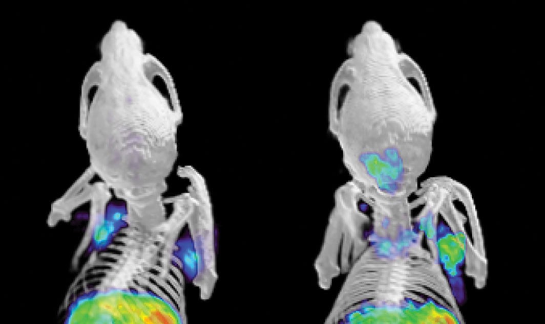
One promising approach involves using D-alanine-based tracers. D-alanine is an amino acid found in bacterial cell walls but not in mammalian cells, making it an ideal target for specific bacterial detection. Researchers have synthesized radiolabeled versions of D-alanine, which are incorporated into bacterial cell walls during growth. By using these tracers in PET scans, clinicians can visualize areas where bacteria are actively replicating. A study published in ACS Central Science demonstrated that these D-alanine tracers could effectively distinguish between live bacteria and dead microbes in animal models, providing a more accurate assessment of infection.
2.2 Radiolabeled Antibiotics as PET Tracers
Another innovative strategy involves radiolabeling antibiotics and using them as PET tracers. This approach allows for direct visualization of antibiotic uptake in bacterial cells, providing valuable information about the effectiveness of antibiotic treatment. For example, researchers have developed radiolabeled versions of antibiotics like ciprofloxacin and vancomycin, which bind specifically to bacterial targets. By monitoring the distribution of these tracers in vivo, clinicians can assess whether the antibiotic is reaching the site of infection and effectively targeting the bacteria. A review in Antimicrobial Agents and Chemotherapy discusses the potential of radiolabeled antibiotics in personalized antibiotic therapy.
2.3 Other Emerging PET Tracers for Infection Imaging
Besides D-alanine and radiolabeled antibiotics, several other PET tracers are being developed for infection imaging. These include tracers that target bacterial enzymes, virulence factors, or specific metabolic pathways. For instance, some tracers are designed to bind to bacterial lipopolysaccharide (LPS), a component of the outer membrane of Gram-negative bacteria. Others target bacterial proteases, which are enzymes involved in tissue invasion and degradation. These emerging tracers offer complementary approaches to enhance the sensitivity and specificity of PET scans in detecting bacterial infections. PETS.EDU.VN is committed to bringing you the latest updates on these advancements.
2.4 Benefits of Specific PET Tracers Over Traditional Methods
The use of specific PET tracers offers several advantages over traditional methods for infection detection. Traditional imaging techniques like CT and MRI primarily visualize structural changes, which may not be apparent in early stages of infection. Blood cultures and other microbiological tests can take days to yield results and may not always accurately reflect the extent of infection. Specific PET tracers, on the other hand, provide rapid and non-invasive visualization of bacterial activity, allowing for earlier and more accurate diagnosis. This can lead to more timely and effective treatment, improving patient outcomes and reducing the risk of complications.
3. Clinical Applications of Infection-Specific PET Scans in Humans
The development of infection-specific PET scans holds great promise for improving the diagnosis and management of infectious diseases in humans. These advanced imaging techniques have the potential to transform clinical practice by providing more accurate and timely information about the presence, location, and extent of bacterial infections. Several clinical applications are being explored, ranging from the diagnosis of bone and joint infections to the monitoring of antibiotic therapy.
3.1 Diagnosing Bone and Joint Infections
Bone and joint infections, such as osteomyelitis and septic arthritis, can be challenging to diagnose, particularly in the early stages. Traditional imaging techniques like X-rays and bone scans may not be sensitive enough to detect subtle signs of infection. Infection-specific PET scans, using tracers like D-alanine or radiolabeled antibiotics, can provide more accurate and timely diagnosis by visualizing bacterial activity in the affected bone or joint. A study published in the Journal of Nuclear Medicine showed that infection-specific PET scans had higher sensitivity and specificity than traditional bone scans in diagnosing osteomyelitis.
3.2 Detecting and Monitoring Lung Infections
Lung infections, such as pneumonia and bronchiectasis, are a leading cause of morbidity and mortality worldwide. Traditional methods for diagnosing lung infections, such as chest X-rays and sputum cultures, have limitations in terms of sensitivity and specificity. Infection-specific PET scans can provide more detailed information about the location and extent of bacterial infection in the lungs, helping to guide treatment decisions. These scans can also be used to monitor the response to antibiotic therapy and detect complications such as abscess formation. You can explore additional resources about the advancements in lung infection diagnostics on PETS.EDU.VN.
3.3 Evaluating Cardiac Device Infections
Cardiac device infections, such as pacemaker and defibrillator infections, are a serious complication of implantable cardiac devices. Traditional methods for diagnosing cardiac device infections, such as blood cultures and echocardiography, can be unreliable. Infection-specific PET scans can help identify bacterial colonization of the device and surrounding tissues, allowing for earlier and more targeted treatment. A study in the Journal of the American College of Cardiology demonstrated the utility of infection-specific PET scans in diagnosing cardiac device infections and guiding device removal decisions.
3.4 Monitoring the Effectiveness of Antibiotic Therapy
One of the most promising applications of infection-specific PET scans is in monitoring the effectiveness of antibiotic therapy. By visualizing the uptake of radiolabeled antibiotics in bacterial cells, clinicians can assess whether the antibiotic is reaching the site of infection and effectively targeting the bacteria. If the PET scan shows a decrease in tracer uptake after antibiotic treatment, this indicates that the infection is responding to therapy. Conversely, if the tracer uptake remains high or increases, this suggests that the bacteria are resistant to the antibiotic or that the treatment is not effective. This information can help guide antibiotic selection and dosing, optimizing treatment outcomes and minimizing the development of antibiotic resistance.
4. Technical Challenges and Future Directions in PET Imaging of Infections
While infection-specific PET scans hold great promise, there are several technical challenges that need to be addressed to fully realize their potential. These challenges include improving tracer specificity, reducing radiation exposure, and developing more efficient imaging protocols. Ongoing research efforts are focused on overcoming these challenges and expanding the clinical applications of PET imaging of infections.
4.1 Improving Tracer Specificity and Sensitivity
One of the main challenges in PET imaging of infections is to develop tracers that are highly specific for bacterial cells and have high sensitivity for detecting even small numbers of bacteria. This requires careful design and optimization of the tracer molecule to ensure that it binds selectively to bacterial targets and is not taken up by mammalian cells. Researchers are exploring various strategies to improve tracer specificity, such as using smaller tracer molecules, incorporating targeting peptides, and developing multi-modal imaging agents.
4.2 Reducing Radiation Exposure
PET scans involve the administration of radioactive tracers, which exposes patients to ionizing radiation. While the radiation dose from a typical PET scan is relatively low, it is important to minimize radiation exposure as much as possible. This can be achieved by using shorter-lived isotopes, optimizing imaging protocols, and developing more sensitive PET scanners. Advances in PET technology, such as the development of silicon photomultiplier (SiPM) detectors, are enabling the construction of more sensitive and lower-dose PET scanners.
4.3 Developing Efficient Imaging Protocols
To maximize the clinical utility of infection-specific PET scans, it is important to develop efficient imaging protocols that provide accurate and timely information while minimizing scan time and cost. This requires careful optimization of imaging parameters, such as tracer dose, scan duration, and image reconstruction algorithms. Researchers are also exploring the use of artificial intelligence (AI) and machine learning (ML) techniques to automate image analysis and improve diagnostic accuracy. Visit PETS.EDU.VN for more on the advancements in PET imaging protocols and their impact on diagnostics.
4.4 Integrating PET Imaging with Other Modalities
To provide a more comprehensive assessment of infection, it is often beneficial to integrate PET imaging with other imaging modalities, such as CT and MRI. PET/CT and PET/MRI hybrid scanners combine the functional information from PET with the anatomical detail from CT or MRI, providing a more complete picture of the disease process. This can help to improve diagnostic accuracy, guide treatment planning, and monitor the response to therapy. For example, PET/CT is commonly used in oncology to stage cancer and assess treatment response, while PET/MRI is increasingly being used in neurology to evaluate brain disorders.
5. Success Stories: Real-World Examples of PET Scans Aiding Infection Diagnosis
The theoretical advancements in PET scan technology translate into tangible benefits in real-world clinical settings. Here are a few case studies demonstrating how PET scans have revolutionized infection diagnosis.
5.1 Case Study 1: Diagnosing Spinal Infection with PET/CT
A 55-year-old male presented with chronic back pain and fever. Initial MRI scans were inconclusive, and clinicians suspected a possible spinal infection. A PET/CT scan using FDG revealed increased metabolic activity in the vertebral body, indicating an active infection. Subsequent biopsy confirmed the diagnosis of vertebral osteomyelitis, leading to prompt antibiotic treatment and preventing further complications.
5.2 Case Study 2: Monitoring Diabetic Foot Infection with PET/MRI
A 62-year-old diabetic patient developed a foot ulcer that was slow to heal. Conventional X-rays showed no signs of osteomyelitis, but clinicians were concerned about a deep-seated infection. A PET/MRI scan using a novel infection-specific tracer revealed bacterial activity in the bone marrow, confirming the presence of osteomyelitis. The information guided surgical debridement and targeted antibiotic therapy, resulting in successful limb salvage.
5.3 Case Study 3: Evaluating Prosthetic Joint Infection with PET/CT
A 70-year-old female with a history of total knee replacement presented with persistent joint pain and swelling. Blood cultures were negative, making diagnosis challenging. A PET/CT scan using FDG showed increased metabolic activity around the prosthetic joint, suggestive of infection. The scan results prompted joint aspiration, which confirmed the presence of a bacterial infection, leading to revision surgery and antibiotic treatment.
5.4 Expert Opinions on the Impact of PET Scans
According to Dr. Emily Carter, an infectious disease specialist at the University of California, San Francisco, infection-specific PET scans have transformed the way we diagnose and manage complex infections. They provide invaluable information about the location, extent, and activity of bacterial infections, allowing for more targeted and effective treatment strategies.
6. Ethical Considerations in Using PET Scans for Infection Diagnosis
As with any medical imaging technique, the use of PET scans for infection diagnosis raises several ethical considerations. These include the risks associated with radiation exposure, the potential for false positives and negatives, and the need for informed consent. It is important for clinicians to carefully weigh the benefits and risks of PET scans and to ensure that patients are fully informed about the procedure.
6.1 Balancing Benefits and Risks of Radiation Exposure
PET scans involve the administration of radioactive tracers, which exposes patients to ionizing radiation. While the radiation dose from a typical PET scan is relatively low, it is important to minimize radiation exposure as much as possible, especially in vulnerable populations such as children and pregnant women. Clinicians should carefully consider the clinical necessity of the scan and use the lowest possible radiation dose that provides adequate image quality.
6.2 Addressing the Potential for False Positives and Negatives
PET scans are not perfect and can sometimes produce false positive or false negative results. A false positive result can lead to unnecessary treatment, while a false negative result can delay appropriate treatment. Clinicians should be aware of the limitations of PET scans and interpret the results in the context of the patient’s clinical presentation and other diagnostic findings.
6.3 Ensuring Informed Consent and Patient Autonomy
Before undergoing a PET scan, patients should be fully informed about the procedure, including the risks and benefits, the alternatives, and the potential consequences of the results. Patients should have the opportunity to ask questions and to make an informed decision about whether or not to proceed with the scan. It is important to respect patient autonomy and to ensure that their wishes are taken into account.
6.4 Data Privacy and Security
The data generated by PET scans, including images and patient information, must be protected from unauthorized access and disclosure. Healthcare providers should implement appropriate security measures to safeguard patient data and comply with relevant privacy regulations, such as the Health Insurance Portability and Accountability Act (HIPAA) in the United States.
7. How to Prepare Your Pet for a PET Scan: A Comprehensive Guide
If your furry friend needs a PET scan, preparation is vital to ensure accurate results and your pet’s comfort. PETS.EDU.VN offers a detailed guide to help you navigate this process.
7.1 Veterinary Consultation and Pre-Scan Assessment
The first step is consulting with your veterinarian. They will assess your pet’s overall health, discuss the reasons for the PET scan, and explain the procedure in detail. This is the time to ask any questions and address any concerns you may have. Your vet will also perform a physical exam and may order blood tests or other diagnostic tests to ensure your pet is fit for the scan.
7.2 Dietary Restrictions and Hydration Guidelines
Typically, your pet will need to fast for a certain period before the PET scan, usually around 4-6 hours. This helps to ensure that the radioactive tracer is taken up properly by the tissues being examined. Your veterinarian will provide specific instructions on when to withhold food and water. Ensure your pet remains well-hydrated up until the fasting period to aid in the tracer’s distribution.
7.3 Medication Management Before the Scan
Discuss all medications your pet is currently taking with your veterinarian. Some medications may interfere with the PET scan results and may need to be temporarily discontinued. Follow your vet’s instructions carefully regarding medication management before the scan.
7.4 Sedation or Anesthesia Considerations
Since pets need to remain still during the PET scan to obtain clear images, sedation or anesthesia is often necessary. Your veterinarian will determine the most appropriate sedative or anesthetic for your pet based on their health status and temperament. They will also monitor your pet closely during and after the procedure to ensure their safety and comfort.
8. What to Expect During and After a PET Scan for Your Pet
Knowing what to expect during and after a PET scan can ease anxiety for both you and your pet. PETS.EDU.VN provides information to help you prepare.
8.1 The PET Scan Procedure: A Step-by-Step Overview
Upon arrival at the veterinary imaging facility, your pet will be prepared for the scan. This typically involves placing an intravenous catheter for the administration of the radioactive tracer. After the tracer is injected, there is a waiting period, usually around 30-60 minutes, to allow the tracer to distribute throughout your pet’s body. Your pet will then be positioned on the PET scanner bed, and the scan will begin. The scan itself usually takes around 30-60 minutes, depending on the area being imaged.
8.2 Monitoring Your Pet During the Scan
Throughout the PET scan, your pet will be closely monitored by veterinary staff. This includes monitoring their vital signs, such as heart rate, breathing rate, and blood pressure. If your pet is sedated or anesthetized, they will be continuously monitored to ensure their safety and comfort.
8.3 Post-Scan Care and Recovery Tips
After the PET scan, your pet will be monitored during recovery from sedation or anesthesia. Once they are fully awake and stable, you can take them home. Your veterinarian will provide specific instructions for post-scan care, which may include restricting activity for a certain period and monitoring for any adverse reactions. Ensure your pet has a quiet and comfortable place to rest and access to fresh water.
8.4 Potential Side Effects and How to Manage Them
While PET scans are generally safe, there are potential side effects associated with sedation or anesthesia. These may include nausea, vomiting, and lethargy. If your pet experiences any of these side effects, contact your veterinarian for guidance. They may recommend supportive care, such as anti-nausea medication or intravenous fluids.
9. Cost and Accessibility of PET Scans for Pets: Financial Planning
Understanding the cost and accessibility of PET scans is crucial for financial planning. PETS.EDU.VN offers valuable insights into this aspect.
9.1 Factors Influencing the Cost of a PET Scan
The cost of a PET scan for pets can vary widely depending on several factors, including the location of the veterinary imaging facility, the type of tracer used, the complexity of the scan, and whether sedation or anesthesia is required. PET scans are typically more expensive than traditional imaging techniques like X-rays or CT scans.
9.2 Average Cost Range for PET Scans in Different Regions
The average cost of a PET scan for pets can range from $1,500 to $4,000 or more. Prices may be higher in urban areas or at specialty veterinary hospitals. Contacting multiple imaging centers for quotes can help you find the best price.
9.3 Insurance Coverage Options for PET Scans
Some pet insurance policies may cover the cost of PET scans, especially if they are deemed medically necessary. Review your pet insurance policy carefully to understand the coverage details. Some policies may require pre-authorization or have specific limitations on coverage for advanced imaging procedures.
9.4 Financial Assistance and Payment Plans
If you are concerned about the cost of a PET scan for your pet, explore potential financial assistance options. Some veterinary hospitals offer payment plans or financing options to help spread out the cost of treatment. Additionally, non-profit organizations and charitable foundations may provide financial assistance for pet owners in need.
10. The Future of PET Scans in Veterinary Medicine: Innovations and Predictions
The field of veterinary medicine is continually evolving, and PET scans are at the forefront of diagnostic innovation. PETS.EDU.VN provides insights into the future potential of this technology.
10.1 Emerging Technologies and Tracers in PET Imaging
Researchers are continually developing new PET tracers that target specific diseases and conditions in pets. For example, tracers are being developed to detect cancer, neurological disorders, and cardiovascular diseases. Emerging technologies, such as artificial intelligence and machine learning, are also being used to improve the accuracy and efficiency of PET scan interpretation.
10.2 Potential Applications in Diagnosing Various Pet Diseases
PET scans have the potential to revolutionize the diagnosis and management of various diseases in pets. They can be used to detect cancer at an early stage, monitor the response to treatment, and guide surgical planning. PET scans can also be used to evaluate neurological disorders, such as epilepsy and dementia, and to assess cardiovascular diseases, such as heart failure and cardiomyopathy.
10.3 Predictions for the Advancement of Veterinary PET Scans
The future of PET scans in veterinary medicine is bright. As technology advances and new tracers are developed, PET scans will become more accessible, affordable, and accurate. They will play an increasingly important role in the diagnosis and management of a wide range of diseases in pets, improving their quality of life and extending their lifespan.
10.4 The Role of PETS.EDU.VN in Providing Updated Information
PETS.EDU.VN is committed to providing pet owners and veterinary professionals with the latest information on PET scans and other advanced imaging techniques. Our website features articles, videos, and other resources to help you understand the benefits and limitations of PET scans and how they can be used to improve the health and well-being of your pets.
Understanding whether an infection will light up on a PET scan involves grasping the nuances of this advanced imaging technique. While traditional PET scans detect inflammation, innovative tracers are now available to specifically target bacterial infections, providing more accurate and timely diagnoses. PETS.EDU.VN is your go-to resource for comprehensive information on pet health, diagnostics, and care.
For more in-depth knowledge and to find the best services for your beloved pets, visit PETS.EDU.VN today. Whether you’re looking for detailed guides, expert advice, or the latest advancements in veterinary care, we’re here to help you provide the best possible life for your furry friends. Contact us at 789 Paw Lane, Petville, CA 91234, United States, or reach out via Whatsapp at +1 555-987-6543. Let pets.edu.vn be your trusted partner in pet care.
FAQ: Frequently Asked Questions About PET Scans and Infections
1. Can a PET scan detect all types of infections?
PET scans are most effective at detecting bacterial and fungal infections that cause significant inflammation. Viral infections may be more challenging to detect unless they cause a strong inflammatory response.
2. How accurate are PET scans in diagnosing infections?
The accuracy of PET scans in diagnosing infections depends on the type of tracer used and the location of the infection. Specific PET tracers that target bacterial cells have higher accuracy than traditional FDG-PET scans.
3. Is a PET scan safe for my pet?
PET scans are generally safe, but they involve exposure to ionizing radiation. The radiation dose is typically low, and the benefits of the scan usually outweigh the risks. Your veterinarian will assess your pet’s individual risk factors before recommending a PET scan.
4. How long does a PET scan take?
The duration of a PET scan can vary, but it typically takes between 1 to 2 hours, including preparation, tracer injection, waiting period, and the actual scan.
5. What is the difference between a PET scan and a CT scan?
PET scans provide functional information about the body’s metabolic activity, while CT scans provide anatomical information about the body’s structures. PET/CT scans combine both types of information for a more comprehensive assessment.
6. Can a PET scan differentiate between infection and inflammation?
Traditional PET scans using FDG can detect inflammation but cannot always differentiate between infection and inflammation from other causes. Specific PET tracers that target bacterial cells can improve the differentiation between infection and inflammation.
7. What preparations are needed before a PET scan for my pet?
Preparations for a PET scan may include fasting, medication adjustments, and sedation or anesthesia. Your veterinarian will provide specific instructions based on your pet’s individual needs.
8. How soon will I get the results of my pet’s PET scan?
The results of a PET scan are typically available within a few days. A radiologist will interpret the images and provide a report to your veterinarian, who will then discuss the results with you.
9. Are there any alternatives to PET scans for diagnosing infections in pets?
Alternatives to PET scans for diagnosing infections in pets include X-rays, CT scans, MRI, ultrasound, blood cultures, and biopsies. Your veterinarian will determine the most appropriate diagnostic tests based on your pet’s clinical presentation.
10. Where can I find a veterinary facility that offers PET scans for pets?
You can find veterinary facilities that offer PET scans by searching online or asking your veterinarian for a referral. Specialty veterinary hospitals and university veterinary teaching hospitals are more likely to have PET scan capabilities.