Positron Emission Tomography (PET) scans are powerful diagnostic tools that provide detailed insights into the body’s functions at a cellular level. If your healthcare provider has recommended a PET scan, understanding the results can be empowering. While the reports can seem dense with medical terms, breaking down key concepts can make them much clearer. This guide focuses on demystifying one of the most crucial aspects of PET scan interpretation: FDG uptake.
While this information is designed to be helpful, it is essential to remember that it should not replace a consultation with your doctor. They are best equipped to interpret your specific results in the context of your overall health. Many providers will schedule a follow-up appointment to discuss your PET scan findings. By familiarizing yourself with terms like FDG uptake beforehand, you can participate more actively in these conversations, ask informed questions, and better understand your health journey. Tools like PocketHealth can also be invaluable, offering secure access to your imaging results as soon as they are available, allowing you to be prepared and informed.
What is a PET Scan?
PET stands for Positron Emission Tomography. It’s a type of nuclear medicine imaging that utilizes a radioactive tracer to visualize the metabolic activity of your tissues and organs. During a PET scan, a small amount of a radioactive substance, known as a radiotracer, is introduced into your body, usually by injection. This tracer emits positrons, which are detected by the PET scanner. The scanner then converts these signals into detailed, color-coded images that reveal how your body’s tissues are functioning.
PET scans are frequently used alongside other imaging techniques such as CT (Computed Tomography) or MRI (Magnetic Resonance Imaging). This combination allows healthcare professionals to assess both the structure and function of organs and tissues, providing a more comprehensive diagnostic picture than either test alone. PET scans vs MRI vs CT scans offer different but complementary information, making them powerful tools in modern medicine.
Axial, Sagittal, and Coronal Planes in PET Scans
To provide a complete view of your internal structures, PET scans capture images in three different planes:
- Axial Plane (Transverse View): This horizontal view divides the body into top and bottom sections, like looking down at a stack of pancakes. It’s crucial for seeing cross-sections of organs.
- Sagittal Plane (Side View): This vertical view divides the body into left and right halves, offering a profile perspective. It’s useful for visualizing structures along the body’s length.
- Coronal Plane (Frontal View): This vertical view divides the body into front and back sections, similar to looking at someone face-to-face. It provides a view of structures from front to back.
Understanding these planes helps you visualize how the 2D images on a PET scan report represent the 3D structures within your body. Radiologists use these different perspectives to get a 360° view and accurately interpret the scan.
Whole body PET scan showing the axial, sagittal and coronal views
The Role of Radiotracers in PET Scans
A radiotracer is a key component of a PET scan. It’s a specially designed substance that emits positrons and is used to track metabolic activity within the body. The most common radiotracer used in PET scans is Fluorodeoxyglucose (FDG). Radiotracers are administered to the patient via injection, inhalation, or ingestion, depending on the specific tracer and the area being examined. Once in the body, the radiotracer circulates and accumulates in tissues and organs based on their metabolic activity.
Your PET scan report will typically specify which radiotracer was used, the amount administered, the administration route, and the location of administration. This information is important for accurate interpretation of the scan results.
Decoding FDG and FDG Uptake in PET Scans
FDG, or Fluorodeoxyglucose, is a glucose analogue, meaning it’s similar to glucose (sugar) but with a radioactive fluorine atom attached. Since glucose is the primary source of energy for cells, especially metabolically active cells, FDG is readily taken up by these cells. This makes FDG an excellent radiotracer for PET scans, particularly for detecting conditions characterized by altered glucose metabolism, such as cancer, infection, and inflammation.
When you see terms like “FDG uptake” or “FDG activity” in your PET scan report, they refer to how much FDG has been absorbed by your body’s cells. Understanding FDG uptake is central to interpreting your PET scan results and gaining insights into your metabolic health.
What Exactly is FDG Uptake?
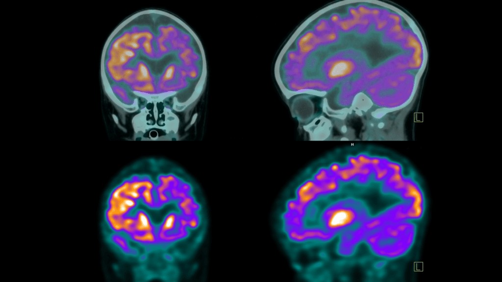 FDG uptake on PET scans
FDG uptake quantifies the amount of FDG radiotracer absorbed by cells in specific areas of the body. Different types of cells have varying metabolic rates, leading to different levels of FDG uptake. Tissues with high metabolic activity, like the brain, heart, and rapidly dividing cancer cells, will typically exhibit higher FDG uptake.
FDG activity and FDG uptake are often used interchangeably, but there’s a subtle distinction. FDG uptake is the measurement of the amount of FDG in the tissue, while FDG activity describes the metabolic processes driving that absorption. Higher FDG uptake generally indicates higher metabolic activity. Think of it like this: FDG uptake is the “what” – the amount of tracer present, and FDG activity is the “why” – the underlying metabolic rate causing that uptake.
Now that you understand the basics of FDG, let’s explore common terms related to FDG uptake that you might encounter in your PET scan report.
No FDG Uptake: Understanding the Implications
“No FDG uptake” means that the cells in a particular area are not absorbing a detectable amount of the FDG radiotracer. The significance of this finding depends heavily on the type of tissue being examined and the clinical context.
Here are some potential interpretations of “no uptake”:
- Inactive Tumor or Growth: If a PET scan is performed to monitor a known tumor, “no uptake” could be a positive sign. It might indicate that the tumor is no longer metabolically active, possibly due to successful treatment, or that the tissue is necrotic (dead). However, further tests are usually needed to confirm this.
- Normal Tissue with Low Metabolic Rate: Some tissues naturally have low metabolic activity and therefore exhibit minimal FDG uptake. In these cases, “no uptake” is a normal and healthy finding.
- Technical Issues: In rare instances, “no uptake” could be due to technical problems with the PET scanner’s sensitivity or functionality.
- Resolved Inflammation or Infection: If the scan was intended to evaluate inflammation or infection, “no uptake” might suggest that the condition has resolved or is currently inactive.
Normal FDG Uptake: Expected Metabolic Activity
Certain organs and tissues, such as the brain, liver, spleen, and bone marrow, are naturally metabolically active and typically show “normal FDG uptake.” This indicates that these tissues are functioning within expected metabolic parameters. “Normal uptake” serves as a baseline, and radiologists are trained to recognize typical FDG distribution patterns throughout the body.
The expected level of “normal FDG uptake” varies depending on the specific tissue type and location. Your PET scan report will be interpreted in the context of these established medical baselines.
Mild or Low FDG Uptake: When to Investigate Further
“Mild” or “low FDG uptake” (sometimes described as “low-level” or “low-grade”) suggests a lower than expected level of metabolic activity in a particular area. While it can be normal for tissues with inherently lower metabolic rates, it can also warrant further investigation in areas where higher activity is typically expected.
Possible interpretations of “mild or low FDG uptake” include:
- Reduced Tissue Viability or Activity: This could indicate tissue necrosis or a decrease in metabolic activity in previously active areas. It might be a sign that a tumor, inflammation, or disease is becoming less active, potentially indicating successful treatment. However, further evaluation is usually necessary.
- Certain Tumor Types: Some types of tumors naturally exhibit lower FDG uptake, even when active.
- Technical Limitations: As with “no uptake,” technical issues with the scanning equipment could sometimes contribute to falsely low readings.
Increased FDG Uptake: Potential Indicators
“Increased FDG uptake” or “intense FDG uptake” signifies that cells in a specific region are absorbing more FDG than the surrounding tissues. This is often visualized as brighter or more intense spots on the PET scan images. “Increased FDG uptake” is a significant finding that requires careful evaluation.
Possible causes of “increased FDG uptake” include:
- Cancer: Cancer cells are characteristically highly metabolic and consume glucose at a higher rate than normal cells, resulting in increased FDG uptake.
- Inflammation and Infection: Areas of active inflammation or infection also exhibit increased FDG uptake due to the heightened metabolic activity of immune cells and tissue repair processes.
- Tissue Healing: Tissues undergoing repair after surgery, injury, or radiation therapy can show temporarily increased FDG uptake.
- Benign Conditions: Certain non-cancerous conditions, such as benign tumors, active thyroid nodules, or some inflammatory conditions, can also demonstrate increased FDG uptake.
It’s crucial to understand that while “increased FDG uptake” can be concerning for cancer, it is not always indicative of malignancy. A radiologist’s expert interpretation, considering your clinical history and other diagnostic findings, is essential for accurate diagnosis.
Abnormal FDG Uptake: Deviation from the Norm
“Abnormal FDG uptake” is a general term indicating that glucose absorption is not within the expected range for the tissue being assessed. This can encompass both lower-than-expected uptake and higher-than-expected uptake. In the case of higher FDG uptake, further investigation is almost always warranted.
Possible implications of “abnormal FDG uptake” mirroring “increased FDG uptake” are:
- Potential Cancer: Many cancers are associated with increased metabolic activity and thus higher FDG uptake.
- Infection or Inflammation: Immune responses to infection or inflammation also lead to elevated metabolic activity and increased FDG uptake.
Standardized Uptake Value (SUV): Quantifying FDG Uptake
SUV, or Standardized Uptake Value, is a quantitative measure used in PET scans to assess the degree of radiotracer uptake in a specific area. It’s a ratio that compares the radiotracer activity in a region of interest to the injected dose and the patient’s body size. SUV provides a numerical value that helps standardize the interpretation of FDG uptake across different patients and scans.
A higher SUV generally indicates greater metabolic activity, which, as discussed earlier, can be associated with various conditions. Typical SUV ranges are often categorized as:
- Low Intensity: SUV < 5
- Moderate: SUV 5-10
- Intense: SUV 10-15
- Very Intense: SUV > 15
SUV is particularly valuable for comparing PET scans performed at different times, such as to monitor treatment response. Changes in SUV over time can provide objective evidence of disease progression or regression.
Physiological Uptake: Normal Biological Activity
“Physiological uptake” refers to the expected and normal absorption of a radiotracer by various organs and tissues due to their natural biological functions. It’s crucial to distinguish physiological uptake from abnormal uptake that might indicate disease. While “FDG uptake” can refer to both normal and abnormal tracer accumulation, “physiological uptake” specifically highlights the normal, expected patterns.
Different organs have different levels of physiological uptake based on their metabolic rates and functions. For example, the brain and heart typically exhibit high physiological uptake due to their high glucose demands. Understanding these normal patterns is key to identifying truly abnormal findings on a PET scan.
Physiological Uptake vs. Cancer: Clearing Up Misconceptions
“Physiological uptake” itself does not mean cancer. It’s a normal phenomenon reflecting the body’s healthy functioning. It’s important not to misinterpret physiological uptake as a sign of disease. Radiologists are trained to differentiate between physiological uptake patterns and abnormal uptake that may be suspicious for cancer or other pathologies.
Physiological Activity in the Liver: A Normal Finding
“Physiological activity in the liver” is a common and normal finding on PET scans. The liver is a highly metabolic organ involved in numerous bodily functions, including glucose metabolism. Therefore, it naturally absorbs FDG radiotracer.
Key points about physiological activity in the liver:
- Normal Expectation: Liver uptake is expected and indicates normal liver function in terms of glucose metabolism.
- Non-Specific: It does not point to any specific disease or abnormality.
- Variable Levels: The degree of physiological uptake can vary slightly based on factors like diet and blood sugar levels, but it remains within a normal range.
Physiological Activity in Kidneys and Bladder: The Excretory System at Work
“Physiological uptake in the kidneys and bladder” is also a normal finding related to the body’s excretion of the radiotracer.
- Kidneys: The kidneys filter waste products from the blood, including the FDG radiotracer. This filtration process leads to physiological uptake in the kidneys as they process and remove the tracer.
- Bladder: The filtered radiotracer is then excreted in urine and collects in the bladder. The bladder will show physiological uptake as it stores the radiotracer before elimination from the body.
Physiological uptake in the kidneys and bladder is a sign that these organs are functioning correctly in processing and eliminating waste, including the radiotracer. It’s not indicative of any disease process.
Metabolic Activity on a PET Scan: A Broader Perspective
“Metabolic activity” on a PET scan is a broad term referring to the rate at which cells are using energy, typically measured by glucose consumption. PET scans, using FDG, directly visualize this metabolic activity, highlighting areas with increased or decreased glucose uptake. Understanding metabolic activity is central to interpreting PET scan findings.
No Metabolic Activity: Potential Interpretations
“No metabolic activity” can have different meanings depending on the context. While in some tissues, it’s normal, in others, it can be significant.
Potential implications of “no metabolic activity” as an abnormal finding include:
- Tissue Damage (Necrosis): Damaged or necrotic tissue may no longer be metabolically active and thus show no FDG uptake.
- Blockages: Reduced blood flow or vascular blockages, such as in stroke or heart attack, can lead to a lack of metabolic activity in the affected area.
- Successful Treatment: In the context of cancer treatment, “no metabolic activity” in a previously active tumor can be a very positive sign, indicating successful treatment and tumor response.
Low-Grade or Mild Metabolic Activity: Possible Scenarios
“Low-grade metabolic activity” or “mild metabolic activity” suggests a reduced level of glucose metabolism. As with “no metabolic activity,” its significance varies by tissue type and clinical situation.
Potential interpretations of “low-grade metabolic activity” as an abnormal finding:
- Early Stage Disease: It could represent the early stages of infection or disease, including tumors, where metabolic activity is present but not yet highly elevated.
- Scar Tissue: Scar tissue and fibrous tissues typically have lower metabolic demands compared to healthy or diseased tissues.
- Age-Related Changes: Metabolic rate can naturally decline with age, making low-grade activity a normal finding in some older individuals.
- Metabolic Disorders: Certain metabolic disorders can affect glucose metabolism and FDG uptake.
- Treatment Response: Decreased metabolic activity can also indicate that a treatment is working and reducing the activity of a tumor or inflammation.
Increased Metabolic Activity or Hypermetabolic: A Key Indicator
“Increased metabolic activity” or “hypermetabolic” indicates a higher than normal rate of glucose metabolism in cells. This is a significant finding on PET scans and often warrants further investigation.
“Increased metabolic activity” can be associated with:
- Cancerous Tumors: Rapidly proliferating cancer cells have high glucose demands.
- Inflammation and Infection: Immune cells and inflammatory processes are metabolically active.
- Tissue Repair: Healing tissues require increased energy and can show increased metabolic activity.
- Benign Conditions: Some non-cancerous conditions can also be hypermetabolic.
While “increased metabolic activity” raises concern for serious conditions, it’s not always cancer. Clinical context and further diagnostic workup are crucial for accurate interpretation.
Deauville Score: Standardizing Lymphoma Response Assessment
The Deauville score (DS) is a standardized scoring system specifically used to assess FDG uptake in PET scans for Hodgkin’s and non-Hodgkin’s lymphomas, particularly in treatment response evaluation. It’s a visual scale that compares FDG uptake in lymphoma lesions to physiological uptake in the liver and mediastinum (the space between the lungs).
The Deauville score ranges from 1 to 5:
- No uptake: No FDG uptake in the lesion.
- Slight uptake: Uptake in the lesion is less than or equal to uptake in the mediastinum.
- Uptake above mediastinum, but below liver: Uptake in the lesion is greater than mediastinum but less than liver.
- Moderate uptake above liver: Uptake in the lesion is moderately greater than liver uptake.
- Markedly increased uptake: Uptake in the lesion is markedly greater than liver uptake.
Lower Deauville scores (1 and 2) indicate a better response to treatment, often considered complete responses. Scores of 3 are adequate, while 4 and 5 suggest inadequate response.
“Unremarkable” PET Scan Results: A Positive Outcome
In medical reports, “unremarkable” is generally good news. In the context of a PET scan, “unremarkable” means that the scan did not reveal any significant or abnormal findings. An “unremarkable” PET scan report is what you hope to see, indicating no evidence of disease or abnormality in the areas examined.
Leveraging PocketHealth Report Reader for PET Scan Understanding
PocketHealth empowers patients to access, manage, and understand their medical imaging records, including PET scans. PocketHealth provides rapid and easy access to your PET scan images and reports, often before your follow-up appointment, putting you in control of your health information.
The Report Reader feature within PocketHealth is particularly helpful for deciphering medical terminology. It provides definitions for medical terms directly within your report, making complex language more accessible and understandable. As one patient, Amy J., shared, “With Report Reader, I get explanations for words [in my report] that I don’t understand. I can now discuss my health with my family doctor with more confidence and have a better understanding of what is going on.”
The Value of Understanding Your PET Scan Results
PET scans are invaluable tools for diagnosing and monitoring various diseases. For patients like Jeanne, a breast cancer survivor who needed medical care while abroad, PocketHealth proved to be a crucial resource. Having immediate access to her past imaging allowed her new doctor in Costa Rica to quickly understand her medical history and provide appropriate care, even when far from home.
Understanding your PET scan results empowers you to have more informed and productive conversations with your healthcare provider. Access to your PET scan results through platforms like PocketHealth allows you to be an active participant in your healthcare journey, ask pertinent questions, and make well-informed decisions about your health. By demystifying terms like FDG uptake, you can approach your PET scan results with greater confidence and understanding.