Prostate cancer diagnosis and management have been revolutionized by advancements in medical imaging. Among these, the PSMA PET scan stands out as a highly effective tool for detecting prostate cancer with unprecedented accuracy. But what is a PSMA PET scan, and why is it considered so valuable? This article provides a comprehensive overview of PSMA PET scans, explaining their function, benefits, and role in modern prostate cancer care.
Understanding PSMA PET Imaging for Prostate Cancer
PSMA PET imaging is a cutting-edge diagnostic technique that utilizes Positron Emission Tomography (PET) to visualize prostate cancer cells throughout the body. PSMA, or Prostate-Specific Membrane Antigen, is a protein that is found in significantly higher quantities on the surface of prostate cancer cells compared to normal cells. This characteristic makes PSMA an excellent target for cancer detection.
The procedure involves a special tracer that binds to PSMA. This tracer is radioactive, allowing it to be detected by the PET scanner. When this radiotracer is injected into the patient, it circulates through the body and accumulates in areas where PSMA is present, effectively highlighting prostate cancer cells, even in very small quantities and in locations distant from the prostate gland.
How PSMA PET Scans Work and Their Advantages
PET scans have been used in oncology for many years to detect various types of cancers. However, traditional PET tracers were not particularly effective at imaging prostate cancer. The breakthrough came with the development of new radiotracers, such as piflufolastat F-18 (Pylarify), which are specifically designed to bind to PSMA.
These PSMA-targeted radiotracers work by attaching themselves to the PSMA protein on prostate cancer cells. The radioactive component of the tracer then emits signals that are picked up by the PET scanner. These signals are converted into detailed images, showing the precise locations of prostate cancer cells anywhere in the body.
The primary benefit of PSMA PET scans lies in their superior accuracy compared to older imaging methods. They can detect even minute tumors that might be missed by conventional imaging techniques like CT scans, MRIs, or bone scans. This enhanced sensitivity is crucial for:
- Early Detection: Identifying cancer recurrence at an earlier stage, even when PSA levels are only slightly elevated.
- Accurate Staging: Determining the extent of cancer spread (metastasis) more precisely, which is vital for treatment planning.
- Treatment Monitoring: Assessing how well a patient is responding to therapy by tracking changes in PSMA expression.
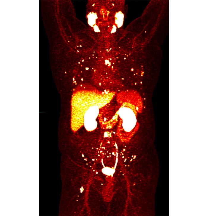 PSMA PET scan showing areas of tumor highlighted as "hot dots".
PSMA PET Scans vs. Traditional Imaging: A Clearer Picture
Traditional imaging methods like CT scans and MRIs rely on anatomical changes to detect tumors, such as changes in tissue density or size. While valuable, these methods are limited in their ability to detect small tumors or cancer at the molecular level. Bone scans, another conventional technique, are used to find metastases in the bones, but they are not as sensitive or specific as PSMA PET scans for prostate cancer.
PSMA PET scans, in contrast, target prostate cancer at a molecular level. By homing in on the PSMA protein, they can reveal the presence of cancer cells long before anatomical changes become visible on CT or MRI. This molecular targeting provides a significant advantage in detecting and characterizing prostate cancer with greater precision and at an earlier stage.
Consider a scenario where a patient has undergone prostatectomy and experiences a rise in PSA levels, suggesting potential recurrence. A CT scan might show a normal lymph node, leading to uncertainty. However, a PSMA PET scan, due to its higher sensitivity, can detect PSMA-expressing prostate cancer cells within that small lymph node, providing crucial information for targeted treatment.
Who is a Good Candidate for a PSMA PET Scan?
PSMA PET imaging is particularly beneficial for specific groups of prostate cancer patients. It is typically recommended for:
- Patients with High-Risk Prostate Cancer: Those whose cancer is likely to have spread beyond the prostate gland. This helps in initial staging to determine if the cancer has metastasized and if it is potentially curable with localized treatments.
- Patients with Suspected Prostate Cancer Recurrence: Individuals who have had prior treatment for prostate cancer (like surgery or radiation) and experience a rise in PSA levels, indicating possible cancer recurrence. PSMA PET scans can help locate the site(s) of recurrence, guiding further treatment.
The decision to undergo a PSMA PET scan should be made in consultation with your physician, who will assess your individual risk factors and medical history.
What to Expect During a PSMA PET Scan Procedure
Undergoing a PSMA PET scan is a relatively straightforward and patient-friendly procedure. No special preparation is typically needed. The process generally involves these steps:
- Radiotracer Injection: Upon arrival at the clinic, you will receive an intravenous injection of the PSMA-targeted radiotracer.
- Waiting Period: You will wait for approximately one hour to allow the radiotracer to circulate throughout your body and be absorbed by any PSMA-expressing cells.
- PET Scan: You will then be positioned in the PET scanner, usually lying down, and remain still for about 20-30 minutes while the machine acquires images. The scan is non-invasive and painless.
- Image Evaluation: The images are typically processed and evaluated by a nuclear medicine physician on the same day. The report and images are then reviewed by your oncology team to guide your treatment plan.
Safety and FDA Approval of PSMA PET Radiotracers
The radiotracer used in PSMA PET scans, piflufolastat F-18 (Pylarify), is FDA-approved and considered safe for diagnostic use. Side effects are rare and generally mild and temporary, such as headache or a temporary alteration in taste.
While the radiotracer is radioactive, the amount of radiation exposure is minimal, comparable to that of a CT scan. The radioactive material also decays rapidly and is eliminated from the body within a few days.
PSMA PET Scan Results and Subsequent Steps
If the PSMA PET scan detects areas of increased tracer uptake, it suggests the presence of prostate cancer. The implications of these findings will depend on your specific clinical situation, including the stage of your cancer and previous treatments.
Based on the scan results, your healthcare team will discuss appropriate treatment options. These may include:
- Targeted Therapies: Treatment directed at specific sites of cancer identified by the PSMA PET scan.
- Systemic Therapies: Body-wide treatments like chemotherapy, hormone therapy, or immunotherapy.
- Theranostic Approaches: In some cases, if the PSMA PET scan is positive, you might be a candidate for theranostic treatments like lutetium-177 PSMA therapy (Pluvicto). This therapy uses a similar PSMA-targeting molecule, but linked to a therapeutic radioactive isotope to deliver radiation directly to cancer cells.
Choosing a Center with Expertise in PSMA PET Scans
Interpreting PSMA PET scans accurately requires specialized expertise in nuclear medicine and prostate cancer imaging. It is essential to choose a medical center with experienced physicians and state-of-the-art technology to ensure the highest quality and most reliable results from your PSMA PET scan. Experienced centers have teams knowledgeable in PSMA PET technology and skilled in interpreting the images to guide optimal patient care.
Accessing PSMA PET Scans
A PSMA PET scan requires a physician’s order. If you believe you might benefit from a PSMA PET scan, discuss it with your doctor. They can determine if it is appropriate for your situation and help you navigate the process of obtaining the scan. Insurance coverage for PSMA PET scans is also increasingly common, but it is advisable to check with your insurance provider to understand your specific coverage details.
In conclusion, what is a PSMA PET scan? It is a revolutionary imaging tool that significantly enhances our ability to detect, stage, and manage prostate cancer. Its superior accuracy and molecular targeting capabilities offer significant advantages over traditional imaging methods, leading to improved patient outcomes and more personalized prostate cancer care.