When your veterinarian recommends a PET scan for your beloved pet, it’s natural to feel a mix of concern and curiosity. Deciphering medical reports, especially those filled with technical terms and acronyms, can be daunting. This guide aims to clarify some common terms you might encounter, specifically focusing on FDG activity in PET scans, to help you better understand your pet’s report and engage in informed discussions with your vet.
While this information is valuable for pet owners, it’s crucial to remember that it is not a substitute for professional veterinary interpretation. Your veterinarian is best equipped to explain your pet’s specific results in detail during a consultation. Familiarizing yourself with the basics beforehand, however, can empower you to ask more pertinent questions and participate actively in your pet’s healthcare journey.
PET Scans for Pets: How Do They Work?
PET stands for Positron Emission Tomography. In veterinary medicine, as in human medicine, PET scans are a type of nuclear imaging. This means they utilize a safe, radioactive substance, known as a radiotracer, to create detailed images of your pet’s organs and tissues.
During a PET scan, a radiotracer is administered to your pet, usually through injection. This tracer travels through the body and is absorbed by tissues. The tracer emits positrons, which are detected by specialized cameras as your pet gently moves through the scanner. These recordings are then processed by a computer to generate colorful images that a veterinary radiologist interprets.
PET scans are frequently used in conjunction with other imaging techniques like CT scans or MRIs to provide a comprehensive assessment. While CT and MRI scans excel at showing the structure of organs, PET scans provide unique insights into their function and metabolic activity.
Navigating the PET Scan Views: Axial, Sagittal, and Coronal Planes
Understanding PET scan views: axial, sagittal, and coronal.
To give veterinarians a complete view of your pet’s internal structures, PET scanners capture images in three different planes:
- Axial Plane (Transverse View): Imagine slicing your pet horizontally – this view shows images of the top and bottom sections of the body.
- Sagittal Plane (Side View): This is a vertical view from the side, showing the left and right halves of your pet’s body.
- Coronal Plane (Frontal View): Picture viewing your pet face-on – this plane provides images of the front and back sections of the body.
These different views are essential for the radiologist to get a 360° perspective and accurately assess all areas of interest.
The Radiotracer: FDG Explained
The key to a PET scan’s functional imaging capability is the radiotracer. This substance, often a type of radioactive sugar, is designed to be absorbed by cells that are metabolically active. Fluorodeoxyglucose (FDG) is the most commonly used radiotracer in PET scans, both for humans and pets.
Your pet’s PET scan report will always specify which radiotracer was used, the amount administered, and the method of administration (injection, inhalation, or ingestion).
Decoding FDG and FDG Uptake: The Heart of Your Pet’s PET Scan Report
FDG, as mentioned, stands for Fluorodeoxyglucose. It’s a glucose analog, meaning it behaves similarly to glucose (sugar) in the body. Since glucose is the primary source of energy for cells, especially highly active cells, FDG becomes a valuable tool for imaging metabolic activity.
When you see terms like “FDG uptake” or “FDG activity” in your pet’s PET scan report, they are referring to how much of this FDG radiotracer has been absorbed and utilized by the cells in different tissues. Understanding FDG uptake is crucial for interpreting the results.
What Exactly is FDG Uptake in Pets?
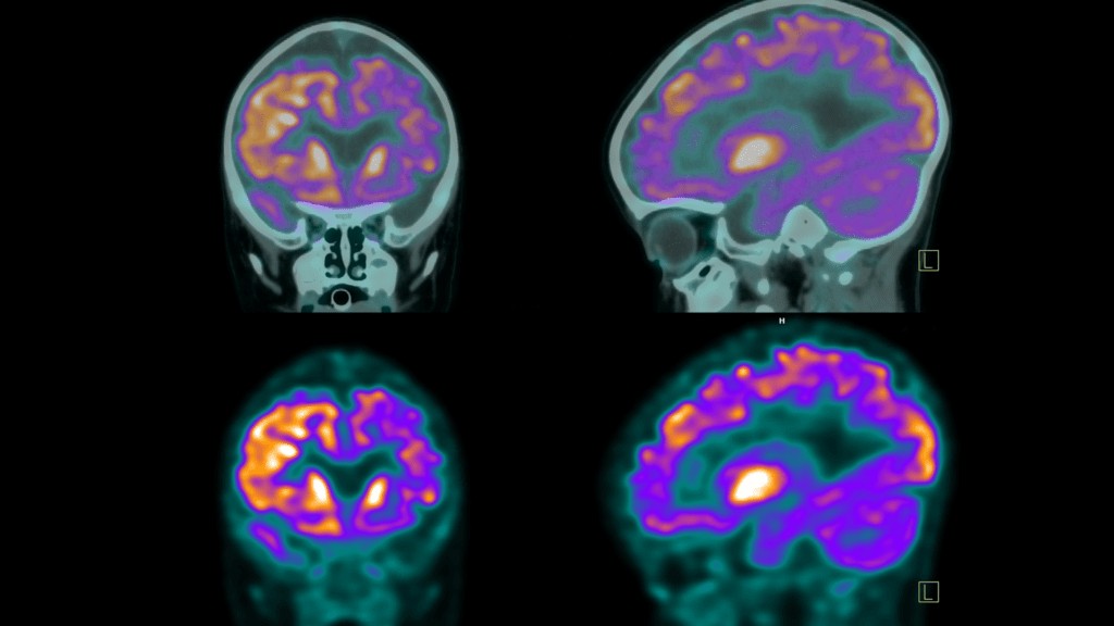 FDG uptake on PET scans
Visualizing FDG uptake in a PET scan.
FDG uptake quantifies the amount of radiotracer absorbed by cells in a specific area. Different types of cells and tissues in your pet’s body have varying metabolic rates and energy requirements. Consequently, FDG will accumulate in different concentrations across the body, highlighting areas of higher metabolic activity.
FDG activity, on the other hand, describes the intensity or vigor with which tissues are using glucose, as reflected by FDG absorption. Higher FDG activity generally indicates higher metabolic activity within those tissues.
In simpler terms: FDG uptake is the measurement of FDG in tissues, while FDG activity describes the metabolic processes causing that absorption.
Now, let’s delve into what different levels of FDG uptake can mean in your pet’s PET scan.
“No Uptake” – What Does it Mean for My Pet?
“No uptake” signifies that FDG is not being absorbed in a particular area. Whether this is a good or bad sign depends heavily on the type of tissue being examined and the clinical context.
- Inactive Tumor or Growth: If your pet has a known tumor or growth, “no uptake” in that area could be a positive sign. It might suggest that the growth is no longer metabolically active, potentially indicating successful treatment or necrosis (tissue death). However, further tests are usually needed for confirmation.
- Normal Tissue: Some tissues naturally have low metabolic activity. In these cases, “no uptake” is considered a normal and healthy result.
- Technical Issues: In rare cases, “no uptake” might be due to technical issues with the scanning equipment or insufficient sensitivity to detect very low levels of uptake.
- Resolved Inflammation/Infection: If the PET scan was performed to evaluate a suspected infection or inflammation, “no uptake” could indicate that the issue has resolved or is not currently active.
“Normal FDG Uptake” – Reassuring Results
Certain organs and tissues in your pet’s body, such as the brain, liver, and spleen, naturally exhibit higher FDG uptake due to their inherently high metabolic demands. For most tissues, there are established “baselines” of expected FDG absorption, which vary by tissue type and location.
“Normal uptake” on your pet’s PET scan report suggests that the tissues are functioning within these expected metabolic ranges. This is generally a reassuring finding.
“Mild” or “Low FDG Uptake” – Context is Key
“Mild” or “low FDG uptake” (sometimes described as “low-level” or “low-grade”) can be normal for tissues with naturally lower metabolic activity, such as fatty tissues. However, in areas expected to be more active, it might warrant closer attention.
- Less Active Tissue: Similar to “no uptake,” “mild uptake” in a previously active area could suggest tissue necrosis or reduced activity of a tumor, inflammation, or disease, potentially indicating successful treatment. Further investigation is often required.
- Tumor Characteristics: Some types of tumors naturally exhibit lower FDG uptake.
- Technical Limitations: As with “no uptake,” technical factors could sometimes play a role.
“Increased FDG Uptake” – A Sign to Investigate
“Increased FDG uptake” or “Intense FDG uptake” means that cells in a specific area are absorbing more FDG radiotracer than the surrounding tissues. This increased uptake appears as brighter or more intense spots on the PET scan images.
In pets, as in humans, increased FDG uptake can be associated with:
- Cancer: Cancer cells are typically highly metabolically active and consume more glucose, leading to increased FDG uptake. This is a primary reason PET scans are valuable in cancer diagnosis and staging.
- Inflammation or Infection: Areas of active inflammation or infection also show increased uptake due to the heightened metabolic activity of immune cells responding to the issue.
- Tissue Healing: Healing tissues after surgery, injury, or radiation therapy may temporarily exhibit increased FDG uptake as part of the repair process.
- Benign Conditions: Certain non-cancerous growths or active thyroid nodules can also show increased FDG uptake.
It’s important to remember that increased FDG uptake does not automatically mean cancer. It indicates increased metabolic activity, which can have various causes, both benign and malignant. Veterinary radiologists and your veterinarian will consider the clinical context, location, and other factors, potentially including further tests, to determine the underlying cause.
“Abnormal FDG Uptake” – Deviation from the Norm
“Abnormal FDG uptake” indicates that glucose absorption is outside the expected range for the specific tissue being assessed. This can mean either lower or higher than normal uptake. In the context of increased abnormal uptake, which is often more concerning, further investigation is usually necessary to determine the cause, with possibilities including cancer, infection, or inflammation, as discussed above.
SUV: Quantifying FDG Uptake
SUV stands for Standardized Uptake Value. It’s a numerical measure used in PET scans to quantify the level of radiotracer activity in a particular area of the image at a specific time. Think of it as a standardized way to measure and compare FDG uptake across different scans and patients (or pets!).
A higher SUV generally suggests increased metabolic activity, while a lower SUV indicates less activity. While specific SUV values are interpreted within the clinical context, general ranges for metabolic activity are often categorized as:
- “Low intensity”: SUV <5
- “Moderate”: SUV 5-10
- “Intense”: SUV 10-15
- “Very intense”: SUV >15
SUV values are particularly helpful for tracking changes over time, such as monitoring a pet’s response to cancer treatment. Changes in SUV between scans can provide valuable information about disease progression or regression.
Physiological Uptake: Normal Metabolic Activity
“Physiological uptake” refers to the expected, normal absorption of a radiotracer in various organs and tissues throughout the body. It’s based on the understanding that different organs naturally have different levels of metabolic activity. While FDG is the most common radiotracer, the concept of physiological uptake applies to any radiotracer.
Understanding physiological uptake patterns allows veterinarians to distinguish between normal metabolic function and potentially abnormal activity that might indicate disease.
Physiological Uptake vs. Cancer: What Pet Owners Need to Know
It’s crucial to understand that physiological uptake does not mean cancer. Normal organs like the brain, heart, liver, kidneys, and bladder naturally take up radiotracers because they are metabolically active. This is expected and normal.
For example, “physiological activity in the liver” on a PET scan simply reflects the liver’s normal metabolic functions, such as glucose metabolism and detoxification. It’s a standard finding and not indicative of disease. Similarly, “physiological activity in the kidneys and bladder” is due to the kidneys filtering the radiotracer and the bladder storing urine containing the tracer – again, normal bodily functions visualized by the scan.
Veterinary radiologists are trained to differentiate between physiological uptake and abnormal uptake that warrants further investigation.
Metabolic Activity: The Core of PET Scan Interpretation
“Metabolic activity” on a PET scan, in essence, refers to how actively cells are using glucose or other metabolic substrates. PET scans, by using radiotracers like FDG, are designed to detect and visualize this activity, highlighting areas of increased metabolic uptake which can be indicative of various conditions.
“No Metabolic Activity” – Implications for Your Pet
“No metabolic activity” can have different meanings depending on the context. In some tissues, it’s normal. In others, it could suggest:
- Damaged Tissue: Tissue may be necrotic or severely damaged, no longer metabolizing effectively.
- Blockages: Reduced blood flow or blockages could be affecting metabolic activity in the area.
- Successful Treatment: In the context of tumors, it could indicate a positive response to treatment, where the tumor is no longer metabolically active.
“Low-Grade Metabolic Activity” – A Range of Possibilities
“Low-grade metabolic activity” (or “mild metabolic activity”) means reduced radiotracer absorption. It can be normal in some tissues or indicate:
- Early Stage Conditions: It might represent the early stages of infections or diseases, including tumors, before metabolic activity becomes highly elevated.
- Scar Tissue: Scar tissue has lower metabolic demands.
- Age-Related Changes: Metabolic rate can naturally decline with age.
- Metabolic Disorders: Conditions like diabetes or hypothyroidism can affect metabolic activity.
- Early Treatment Response: It could indicate that a treatment is beginning to be effective in reducing metabolic activity in a diseased area.
“Increased Metabolic Activity” (Hypermetabolic) – A Key Indicator
“Increased metabolic activity” or “hypermetabolic” signifies that cells are more active than normal and are consuming more glucose. This is often a key finding on PET scans and can indicate:
- Cancerous Tumors: Rapidly dividing cancer cells are highly glucose-avid.
- Inflammation and Infection: Active immune responses increase cellular activity.
- Tissue Repair: Healing tissues require increased energy.
- Benign Conditions: Some non-cancerous conditions can also be hypermetabolic.
Increased metabolic activity is a significant finding that requires careful interpretation by veterinary specialists, considering your pet’s overall health picture and potentially requiring further diagnostic steps.
Deauville Score: Specifically for Lymphoma in Pets
The Deauville score (DS) is a scoring system used specifically for evaluating FDG uptake in lymphoma, a common cancer in pets. It’s a visual assessment that compares FDG uptake in affected areas to uptake in normal tissues like the liver and mediastinum (the space in the chest cavity around the heart and major vessels).
The Deauville score ranges from 1 to 5, with lower scores being better:
- No uptake
- Slight uptake, equal to or below mediastinum uptake
- Uptake above mediastinum, but below liver uptake
- Uptake moderately above liver uptake
- Markedly increased uptake compared to liver
In the Deauville scoring system for lymphoma treatment response, scores of 1 and 2 are generally considered complete responses, 3 is adequate, while 4 and 5 suggest inadequate response.
“Unremarkable” – The Best Word in a PET Scan Report
In medical reports, including veterinary PET scan reports, “unremarkable” is actually good news! It means that the PET scan did not reveal any abnormal findings. An “unremarkable” PET scan is what every pet owner hopes to see.
Understanding Your Pet’s PET Scan Results: Partnering with Your Vet
Understanding the terminology in your pet’s PET scan report, especially FDG activity and related concepts, can empower you to have more informed conversations with your veterinarian. While this guide provides a starting point, your vet is your best resource for interpreting your pet’s individual results and developing the most appropriate care plan. Don’t hesitate to ask questions, seek clarification, and work collaboratively with your veterinary team to ensure the best possible health outcomes for your furry companion.