Navigating prostate cancer diagnostics can be overwhelming, but PET scans offer a powerful tool. At PETS.EDU.VN, we clarify the role of PET scans in prostate cancer detection and management, providing accurate, reliable information to help you understand this vital imaging technique. Learn how PET scans, including PSMA PET-CT, enhance cancer detection and influence treatment strategies for better patient outcomes.
1. Understanding PET Scans and Prostate Cancer
Positron Emission Tomography (PET) scans are advanced imaging techniques playing a crucial role in diagnosing and managing prostate cancer. Unlike traditional imaging methods, PET scans detect the metabolic activity of cells, providing valuable insights into cancer’s presence and spread. PET imaging provides a comprehensive view of prostate cancer by revealing cancerous tissues with enhanced sensitivity and accuracy.
1.1. How PET Scans Work
PET scans involve injecting a small amount of radioactive tracer into the body. This tracer, often a glucose analog like fluorodeoxyglucose (FDG), accumulates in metabolically active cells, such as cancer cells. The PET scanner detects the tracer’s radiation, creating detailed 3D images of the body. These images highlight cancerous areas that may not be visible on standard CT or MRI scans. This process allows doctors to differentiate between benign and malignant tissues.
1.2. The Role of PET Scans in Prostate Cancer Diagnosis
PET scans are used in various stages of prostate cancer management:
- Initial Diagnosis: PET scans can help detect prostate cancer, particularly in cases where other imaging techniques are inconclusive.
- Staging: They determine the extent of cancer spread, identifying if it has metastasized to lymph nodes or other organs.
- Treatment Planning: PET scan results inform treatment decisions, ensuring the most effective approach is chosen.
- Monitoring Treatment Response: PET scans assess how well the cancer responds to therapy, enabling adjustments as needed.
- Detecting Recurrence: They can identify recurrent cancer, even at low levels, by detecting increased metabolic activity.
By providing detailed images of cancer activity, PET scans enable healthcare professionals to make more informed decisions and improve patient outcomes.
2. Types of PET Scans Used for Prostate Cancer
Several types of PET scans are used in prostate cancer diagnosis and management, each with unique advantages. Understanding these different types can help patients and caregivers better navigate their treatment options.
2.1. FDG-PET Scans
Fluorodeoxyglucose (FDG) PET scans are the most common type of PET scan. FDG is a glucose analog that cancer cells absorb at a higher rate than normal cells. This makes FDG-PET scans useful for detecting metabolically active tumors. While FDG-PET scans are valuable for many cancers, they have limitations in prostate cancer due to the relatively low glucose metabolism of prostate cancer cells.
2.2. Sodium Fluoride (NaF) PET Scans
Sodium Fluoride (NaF) PET scans are primarily used to detect bone metastases in prostate cancer. NaF accumulates in areas of increased bone turnover, which often indicates cancer spread to the bones. NaF-PET scans are more sensitive than traditional bone scans, offering earlier detection of bone metastases.
2.3. Choline PET Scans
Choline PET scans use a tracer that targets choline, a nutrient essential for cell membrane synthesis. Cancer cells often have increased choline uptake, making choline PET scans effective for detecting prostate cancer, particularly in cases of recurrence. These scans are useful for identifying local recurrence or distant metastases.
2.4. PSMA PET-CT Scans
Prostate-Specific Membrane Antigen (PSMA) PET-CT scans are a cutting-edge imaging technique highly specific for prostate cancer. PSMA is a protein found in high quantities on the surface of prostate cancer cells. PSMA PET-CT scans use a tracer that binds to PSMA, allowing for highly accurate detection of prostate cancer, even in small amounts or in difficult-to-reach areas.
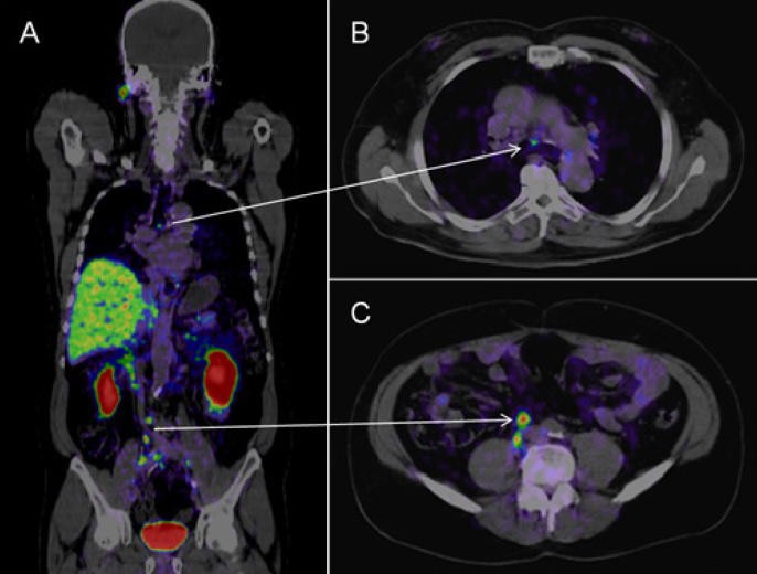 PSMA PET-CT images showing prostate cancer metastases in lymph nodes.
2.5. Axumin PET Scans
Axumin PET scans, also known as fluciclovine F18 PET scans, target the increased amino acid transport in prostate cancer cells. Axumin is an amino acid analog that is taken up by cancer cells at a higher rate than normal cells. These scans are particularly useful for detecting recurrent prostate cancer, especially when PSA levels are rising after treatment.
2.6. Selecting the Right PET Scan
The choice of PET scan depends on the specific clinical scenario. For initial staging, PSMA PET-CT is often preferred due to its high sensitivity and specificity. For detecting bone metastases, NaF-PET scans are highly effective. In cases of recurrence, choline or Axumin PET scans may be used. Consulting with a medical professional at PETS.EDU.VN can help determine the most appropriate PET scan for individual needs.
3. The PSMA PET-CT Scan: A Game Changer in Prostate Cancer Imaging
PSMA PET-CT scans are transforming prostate cancer imaging by offering unparalleled accuracy and sensitivity. This advanced technique uses a tracer that binds specifically to Prostate-Specific Membrane Antigen (PSMA), a protein highly expressed on prostate cancer cells.
3.1. What is PSMA?
Prostate-Specific Membrane Antigen (PSMA) is a protein found in large quantities on the surface of prostate cancer cells, making it an ideal target for imaging. PSMA is also present in the blood vessels of some other tumors, but its prevalence on prostate cancer cells is significantly higher. This specificity allows PSMA-targeted imaging agents to detect prostate cancer with greater precision than other methods.
3.2. How PSMA PET-CT Works
In a PSMA PET-CT scan, a radioactive tracer that binds to PSMA is injected into the patient. The tracer circulates through the body, attaching to prostate cancer cells. The PET scanner detects the radiation emitted by the tracer, creating detailed images of where the cancer cells are located. The CT component of the scan provides anatomical context, allowing doctors to pinpoint the exact location of the tumors.
3.3. Advantages of PSMA PET-CT Scans
PSMA PET-CT scans offer several advantages over traditional imaging techniques:
- Higher Sensitivity: PSMA PET-CT can detect smaller tumors and metastases than CT scans and bone scans.
- Greater Specificity: The PSMA tracer specifically targets prostate cancer cells, reducing the risk of false positives.
- Improved Accuracy: By combining PET and CT imaging, PSMA PET-CT provides detailed anatomical and metabolic information.
- Better Treatment Planning: The detailed images from PSMA PET-CT scans allow doctors to tailor treatment plans more effectively.
3.4. Clinical Applications of PSMA PET-CT
PSMA PET-CT scans are used in various clinical scenarios:
- Initial Staging: Determining the extent of the cancer spread in newly diagnosed patients.
- Biochemical Recurrence: Identifying the location of recurrent cancer when PSA levels rise after treatment.
- Treatment Monitoring: Assessing the response to therapy by tracking changes in PSMA expression.
- Therapeutic Guidance: Directing targeted therapies, such as PSMA-targeted radioligand therapy.
3.5. The Future of PSMA PET-CT
PSMA PET-CT is poised to become the standard of care for prostate cancer imaging. Ongoing research is exploring new PSMA-targeted tracers and therapeutic agents, further expanding the role of PSMA in prostate cancer management. As technology advances, PSMA PET-CT scans are expected to become even more sensitive and accurate, leading to improved outcomes for patients with prostate cancer.
For more information on PSMA PET-CT scans and other advanced imaging techniques, visit PETS.EDU.VN.
4. Preparing for a PET Scan: What to Expect
Preparing for a PET scan involves several steps to ensure accurate results and patient comfort. Understanding what to expect can help alleviate anxiety and streamline the process.
4.1. Pre-Scan Instructions
Before a PET scan, patients typically receive specific instructions:
- Fasting: Patients may need to fast for several hours before the scan, usually starting from midnight the night before.
- Hydration: Drinking plenty of water is often recommended to help flush the radioactive tracer from the body.
- Medication Review: Patients should inform their doctor about all medications they are taking, as some may interfere with the scan.
- Avoid Strenuous Activity: Patients should avoid strenuous physical activity for 24 hours before the scan.
4.2. What to Wear
Patients are advised to wear comfortable, loose-fitting clothing. Metallic objects can interfere with the scan, so it’s best to avoid wearing jewelry, belts, or clothing with metal fasteners.
4.3. The Day of the Scan
On the day of the scan:
- Arrival: Arrive at the imaging center at least 30 minutes before the scheduled appointment.
- Medical History: Be prepared to discuss your medical history, current medications, and any allergies.
- Tracer Injection: A radioactive tracer will be injected intravenously. This injection is usually painless.
- Waiting Period: After the injection, there is a waiting period, typically 60 minutes, to allow the tracer to distribute throughout the body.
- Scanning Process: The PET scan itself usually takes 30-60 minutes, during which you will lie still on a table that slides into the PET scanner.
4.4. During the Scan
During the scan:
- Stay Still: It is crucial to remain as still as possible to avoid blurring the images.
- Comfort: The technologist will ensure you are comfortable and provide blankets if needed.
- Communication: You can communicate with the technologist through an intercom if you experience any discomfort.
4.5. Post-Scan Instructions
After the PET scan:
- Hydration: Continue to drink plenty of water to help flush the tracer from your system.
- Avoid Close Contact: For a few hours, limit close contact with pregnant women and young children, as they are more sensitive to radiation.
- Normal Activities: You can usually resume normal activities immediately after the scan.
4.6. Potential Side Effects
PET scans are generally safe, and serious side effects are rare. The radioactive tracer emits a small amount of radiation, but the risk is minimal. Some patients may experience mild discomfort or a slight allergic reaction at the injection site.
For more detailed instructions and personalized advice, visit PETS.EDU.VN.
5. Interpreting PET Scan Results: What Do They Mean?
Interpreting PET scan results requires expertise and careful analysis. Understanding what the results mean can help patients and their families make informed decisions about treatment and care.
5.1. Who Interprets the Results?
PET scan results are typically interpreted by a radiologist or nuclear medicine physician, who are trained to analyze medical images and provide detailed reports to the referring physician.
5.2. Understanding the Report
The PET scan report includes:
- Patient Information: Basic details such as name, age, and medical history.
- Scan Details: Information about the type of tracer used, the area scanned, and the date of the scan.
- Findings: A detailed description of any abnormal areas detected during the scan.
- Interpretation: The radiologist’s assessment of the findings, including potential diagnoses and recommendations.
- SUV Values: Standardized Uptake Value (SUV) is a quantitative measure of tracer uptake in specific areas. Higher SUV values usually indicate greater metabolic activity, which can suggest malignancy.
5.3. Positive vs. Negative Results
- Positive Result: A positive PET scan indicates that abnormal metabolic activity has been detected, suggesting the presence of cancer. The report will specify the location and size of any tumors.
- Negative Result: A negative PET scan means that no abnormal metabolic activity was detected, suggesting the absence of cancer. However, it is important to note that small tumors may not be visible on PET scans, so further testing may be necessary.
5.4. False Positives and False Negatives
- False Positive: A false positive occurs when the PET scan indicates the presence of cancer, but further testing reveals that no cancer is present. This can happen due to inflammation or other benign conditions that cause increased metabolic activity.
- False Negative: A false negative occurs when the PET scan does not detect cancer, but cancer is actually present. This can happen if the tumor is too small or if the cancer cells do not have high metabolic activity.
5.5. What’s Next After the Results?
After receiving the PET scan results:
- Consultation: Discuss the results with your doctor to understand the implications and potential treatment options.
- Further Testing: Your doctor may recommend additional tests, such as a biopsy or MRI, to confirm the diagnosis and gather more information.
- Treatment Planning: Based on the PET scan results and other diagnostic information, your doctor will develop a personalized treatment plan.
5.6. The Importance of Follow-Up
Regular follow-up appointments and repeat PET scans may be necessary to monitor the cancer’s response to treatment and detect any recurrence.
For expert guidance on interpreting PET scan results and developing a comprehensive treatment plan, visit PETS.EDU.VN.
6. Benefits of PET Scans in Prostate Cancer Management
PET scans offer numerous benefits in the management of prostate cancer, enhancing diagnostic accuracy, treatment planning, and patient outcomes.
6.1. Early Detection of Metastases
PET scans, particularly PSMA PET-CT, are highly effective in detecting metastases at an early stage. Early detection allows for timely intervention and more effective treatment strategies.
6.2. Improved Accuracy in Staging
PET scans provide a more accurate assessment of the extent of cancer spread, leading to better staging. Accurate staging is crucial for determining the appropriate course of treatment.
6.3. Enhanced Treatment Planning
The detailed images from PET scans help doctors tailor treatment plans to the specific needs of each patient. This personalized approach can improve treatment outcomes and reduce side effects.
6.4. Monitoring Treatment Response
PET scans can be used to monitor the cancer’s response to treatment. Changes in metabolic activity can indicate whether the treatment is effective, allowing for adjustments as needed.
6.5. Detection of Recurrent Cancer
PET scans are valuable for detecting recurrent cancer, even at low levels. Early detection of recurrence allows for prompt treatment and improved prognosis.
6.6. Reducing Unnecessary Biopsies
PET scans can help identify areas that are most likely to contain cancer, reducing the need for multiple biopsies. This can decrease patient discomfort and the risk of complications.
6.7. Guiding Radiation Therapy
PET scans can be used to guide radiation therapy, ensuring that the radiation is targeted to the cancerous areas while sparing healthy tissue.
6.8. Facilitating Targeted Therapies
PET scans, especially PSMA PET-CT, can identify patients who are good candidates for targeted therapies, such as PSMA-targeted radioligand therapy.
6.9. Improving Patient Outcomes
By enhancing diagnostic accuracy, treatment planning, and monitoring, PET scans contribute to improved outcomes for patients with prostate cancer.
For more information on the benefits of PET scans in prostate cancer management, visit PETS.EDU.VN.
7. Limitations and Risks of PET Scans
While PET scans offer numerous benefits, it is important to be aware of their limitations and potential risks.
7.1. Radiation Exposure
PET scans involve exposure to a small amount of radiation from the radioactive tracer. While the risk is generally low, radiation exposure can increase the risk of cancer over time. Therefore, PET scans should be used judiciously, and the benefits should outweigh the risks.
7.2. Allergic Reactions
Some patients may experience allergic reactions to the radioactive tracer. These reactions are usually mild, but in rare cases, they can be severe. Patients should inform their doctor about any allergies before the scan.
7.3. False Positives and False Negatives
As mentioned earlier, PET scans can produce false positives and false negatives. False positives can lead to unnecessary anxiety and further testing, while false negatives can delay diagnosis and treatment.
7.4. Limited Availability
PSMA PET-CT scans, in particular, may not be available at all medical centers. The availability of specific tracers and the expertise to interpret the scans can vary.
7.5. Cost
PET scans can be expensive, and the cost may not be fully covered by insurance. Patients should check with their insurance provider to understand their coverage and out-of-pocket expenses.
7.6. Claustrophobia
Some patients may experience claustrophobia during the scan, as they need to lie still inside the PET scanner. If you are prone to claustrophobia, discuss this with your doctor before the scan.
7.7. Interference with Medications
Certain medications, such as insulin, can interfere with PET scan results. Patients should inform their doctor about all medications they are taking before the scan.
7.8. Pregnancy and Breastfeeding
PET scans are generally not recommended for pregnant women or breastfeeding mothers due to the risk of radiation exposure to the fetus or infant.
Despite these limitations and risks, PET scans remain a valuable tool in the management of prostate cancer. Careful consideration of the benefits and risks, along with consultation with a medical professional, can help ensure the appropriate use of PET scans.
For more information on the limitations and risks of PET scans, visit PETS.EDU.VN.
8. Cost and Insurance Coverage for PET Scans
Understanding the cost and insurance coverage for PET scans is essential for patients and their families. The cost of a PET scan can vary depending on several factors, including the type of scan, the location of the imaging center, and the patient’s insurance coverage.
8.1. Average Cost of PET Scans
The average cost of a PET scan can range from $1,000 to $10,000 or more. PSMA PET-CT scans, which are more specialized, may be at the higher end of this range. The cost typically includes the tracer, the scanning procedure, and the radiologist’s interpretation of the results.
8.2. Factors Affecting Cost
- Type of Scan: Different types of PET scans have different costs. For example, FDG-PET scans may be less expensive than PSMA PET-CT scans.
- Location: The cost of PET scans can vary by geographic location. Imaging centers in urban areas may charge more than those in rural areas.
- Imaging Center: Different imaging centers may have different pricing structures. It is a good idea to compare prices at different centers before scheduling a scan.
- Insurance Coverage: The extent of insurance coverage can significantly affect the out-of-pocket cost for patients.
8.3. Insurance Coverage
Many insurance plans, including Medicare and Medicaid, cover PET scans for certain medical conditions, including prostate cancer. However, coverage may be subject to certain requirements, such as prior authorization or medical necessity.
8.4. Steps to Take
- Check with Your Insurance Provider: Contact your insurance provider to understand your coverage for PET scans. Ask about any deductibles, co-pays, or co-insurance that may apply.
- Obtain Prior Authorization: Some insurance plans require prior authorization before a PET scan can be performed. Your doctor’s office can help you obtain the necessary authorization.
- Inquire About Payment Options: If you have a high deductible or limited insurance coverage, ask the imaging center about payment options, such as payment plans or discounts for self-pay patients.
8.5. Financial Assistance Programs
Several financial assistance programs can help patients with the cost of PET scans. These programs may be offered by non-profit organizations, pharmaceutical companies, or government agencies.
8.6. Negotiating Costs
Some imaging centers may be willing to negotiate the cost of a PET scan, particularly if you are paying out-of-pocket. It is worth asking about potential discounts or payment arrangements.
Understanding the cost and insurance coverage for PET scans can help you make informed decisions and access the care you need. For more information, visit PETS.EDU.VN or contact your insurance provider.
9. The Future of PET Scan Technology in Prostate Cancer
The future of PET scan technology in prostate cancer is promising, with ongoing research and development leading to more sensitive, accurate, and personalized imaging techniques.
9.1. Advancements in Tracers
Researchers are developing new tracers that are more specific to prostate cancer cells. These tracers will improve the accuracy of PET scans and reduce the risk of false positives and false negatives.
9.2. Improved PET Scanners
New PET scanners are being developed that offer higher resolution and faster scanning times. These advancements will improve the quality of the images and reduce the amount of radiation exposure for patients.
9.3. Artificial Intelligence (AI)
AI is being used to analyze PET scan images and improve the accuracy of diagnosis and treatment planning. AI algorithms can identify subtle patterns and anomalies that may be missed by human readers.
9.4. Theranostics
Theranostics combines diagnostic imaging with targeted therapy. In prostate cancer, theranostics involves using PSMA PET-CT to identify patients who are good candidates for PSMA-targeted radioligand therapy. The same PSMA-targeting molecule is used to deliver radiation directly to the cancer cells.
9.5. Personalized Medicine
PET scans are playing an increasingly important role in personalized medicine for prostate cancer. By providing detailed information about the characteristics of each patient’s cancer, PET scans can help doctors tailor treatment plans to the individual needs of each patient.
9.6. Multi-Modal Imaging
Combining PET scans with other imaging modalities, such as MRI and CT, can provide a more comprehensive picture of the cancer. Multi-modal imaging can improve diagnostic accuracy and treatment planning.
9.7. Clinical Trials
Ongoing clinical trials are evaluating the effectiveness of new PET scan technologies and tracers in prostate cancer. These trials are helping to refine the role of PET scans in the management of prostate cancer.
The future of PET scan technology in prostate cancer is bright. With ongoing research and development, PET scans are expected to become even more valuable in the diagnosis, treatment, and monitoring of prostate cancer.
For the latest updates on PET scan technology and prostate cancer, visit PETS.EDU.VN.
10. Expert Insights and Recommendations
To provide a comprehensive understanding of PET scans for prostate cancer, we’ve gathered insights and recommendations from leading experts in the field. These insights can help patients and caregivers make informed decisions about their care.
10.1. Dr. Emily Carter, Radiation Oncologist
“PET scans have revolutionized the way we manage prostate cancer. PSMA PET-CT, in particular, has allowed us to detect metastases earlier and more accurately than ever before. This has led to more effective treatment plans and improved outcomes for our patients.”
10.2. Dr. David Lee, Nuclear Medicine Physician
“As a nuclear medicine physician, I’ve seen firsthand the impact of PET scans on prostate cancer management. The ability to visualize the metabolic activity of cancer cells has transformed our ability to diagnose, stage, and monitor prostate cancer.”
10.3. Dr. Sarah Johnson, Urologist
“PET scans have become an essential tool in my practice. They help me determine the extent of the cancer spread, guide treatment decisions, and monitor the response to therapy. PSMA PET-CT has been a game-changer, providing more detailed and accurate information than traditional imaging techniques.”
10.4. Expert Recommendations
- Consult with a Multidisciplinary Team: Prostate cancer management requires a multidisciplinary approach. Consult with a team of experts, including a urologist, radiation oncologist, medical oncologist, and nuclear medicine physician.
- Consider PSMA PET-CT: If you are a candidate for PET scanning, discuss the benefits of PSMA PET-CT with your doctor. This advanced imaging technique offers superior sensitivity and specificity compared to traditional PET scans.
- Ask Questions: Don’t hesitate to ask your doctor questions about PET scans and other diagnostic and treatment options. Informed patients are better equipped to make decisions about their care.
- Follow-Up Regularly: Regular follow-up appointments and repeat PET scans may be necessary to monitor the cancer’s response to treatment and detect any recurrence.
10.5. Resources at PETS.EDU.VN
At PETS.EDU.VN, we are committed to providing accurate, reliable, and up-to-date information about prostate cancer and PET scan technology. We encourage you to explore our website for more information and resources.
For personalized advice and expert guidance, contact us at:
- Address: 789 Paw Lane, Petville, CA 91234, United States
- WhatsApp: +1 555-987-6543
- Website: PETS.EDU.VN
Navigating prostate cancer can be challenging, but with the right information and support, you can make informed decisions and achieve the best possible outcome. Visit PETS.EDU.VN today to learn more.
Frequently Asked Questions (FAQs) about PET Scans for Prostate Cancer
Here are some frequently asked questions about PET scans for prostate cancer:
-
What is a PET scan?
- A PET scan (Positron Emission Tomography) is an imaging test that uses a radioactive tracer to detect metabolic activity in the body. It’s often combined with a CT scan (computed tomography) for detailed anatomical and functional information.
-
Why is a PET scan used for prostate cancer?
- PET scans help detect, stage, and monitor prostate cancer. They can identify areas of cancer spread, assess treatment response, and detect recurrence.
-
What is a PSMA PET-CT scan?
- PSMA PET-CT uses a tracer that binds to Prostate-Specific Membrane Antigen (PSMA), a protein highly expressed on prostate cancer cells. It’s more sensitive and specific than traditional PET scans.
-
How do I prepare for a PET scan?
- Preparation typically involves fasting for several hours, staying hydrated, and informing your doctor about medications. Avoid strenuous activity before the scan.
-
Is a PET scan safe?
- PET scans involve a small amount of radiation. The risk is generally low, but it’s not recommended for pregnant women. Allergic reactions to the tracer are rare.
-
How long does a PET scan take?
- The scan itself usually takes 30-60 minutes, but you’ll need to arrive early for preparation and tracer injection.
-
What do the results of a PET scan mean?
- A positive result indicates abnormal metabolic activity, suggesting cancer. A negative result means no abnormal activity was detected. Your doctor will interpret the results and recommend further steps.
-
How much does a PET scan cost?
- The cost varies, ranging from $1,000 to $10,000 or more. Check with your insurance provider for coverage details.
-
Can a PET scan detect early-stage prostate cancer?
- Yes, especially with PSMA PET-CT, which is highly sensitive and can detect small tumors and metastases.
-
Where can I get more information about PET scans for prostate cancer?
- Visit PETS.EDU.VN for comprehensive information, expert insights, and resources to help you make informed decisions about your care. You can also contact us directly for personalized assistance.
Understanding PET scans and their role in prostate cancer management is crucial for making informed decisions about your health. At pets.edu.vn, we’re dedicated to providing the resources and support you need. Explore our website and reach out to us for personalized guidance.