When your healthcare provider suggests a PET scan, it’s normal to feel a mix of curiosity and perhaps a little apprehension, especially when it comes to understanding the results. Medical reports, particularly those from PET scans, can be filled with unfamiliar terms and acronyms, making it challenging to grasp what they truly mean. One term that frequently appears and can cause confusion is “uptake.” This guide aims to demystify what “uptake” means in a PET scan context, along with other common terms, to help you better understand your report and have more informed conversations with your doctor.
While this information is designed to be helpful, it’s crucial to remember that it is not a substitute for professional medical interpretation. Your doctor is best equipped to review your specific results and discuss them with you in detail. However, by familiarizing yourself with terms like “uptake” beforehand, you can approach your follow-up appointment feeling more prepared and ready to ask insightful questions. Platforms like PocketHealth offer secure and easy access to your imaging results, empowering you to take a more active role in understanding your health.
What is a PET Scan?
PET stands for Positron Emission Tomography. PET scans fall under the category of nuclear medicine procedures because they utilize a radioactive substance, known as a radiotracer, to create detailed images of your body’s organs and tissues at a cellular level. During a PET scan, a small amount of radiotracer is introduced into your body, usually through injection, inhalation, or ingestion. This radiotracer emits positrons, which are detected by specialized cameras as you lie within the PET scanner, a machine that resembles a large donut. These recordings are then processed and compiled into colorful images that radiologists interpret.
PET scans are often used in conjunction with other imaging modalities like CT scans or MRIs to provide a comprehensive assessment. While CT scans and MRIs are excellent for visualizing the structure of organs, PET scans excel at revealing how these organs and tissues are functioning, particularly their metabolic activity. This combination of functional and structural information makes PET scans a powerful diagnostic tool.
Understanding Anatomical Planes: Axial, Sagittal, and Coronal
To provide a comprehensive view of your internal body structures, PET scanners capture images across three distinct planes:
Whole body PET scan showing the axial, sagittal and coronal views
Alt text: PET scan illustration showing axial, sagittal, and coronal planes for comprehensive anatomical views.
- Axial Plane: Also known as the transverse view, this plane provides a horizontal cross-section, effectively dividing the body into top and bottom portions. Imagine looking down at your body from above; this is the perspective of an axial view.
- Sagittal Plane: This is the side view, producing lateral images that divide the body into left and right halves. A sagittal view is like looking at a profile of your body.
- Coronal Plane: Referred to as the frontal view, the coronal plane creates images that divide the body into front (anterior) and back (posterior) sections. This is akin to looking at someone face-on.
These three planes work together to give radiologists a 360-degree perspective of your internal organs and structures, ensuring a thorough evaluation.
The Role of Radiotracers in PET Scans
The magic behind PET scans lies in the radiotracer. This radioactive substance, often a type of sugar molecule, is designed to travel through your body and accumulate in cells that are highly active and require a lot of energy. Fluorodeoxyglucose (FDG) is the most commonly used radiotracer in PET scans.
Your PET scan report will specify which radiotracer was used, the administered amount (dosage), the administration site, and the method of delivery (injection, inhalation, or ingestion). This information is important for accurate interpretation of your scan results.
FDG and FDG Uptake: Key Concepts in PET Scan Interpretation
FDG, short for Fluorodeoxyglucose, is a crucial radiotracer that allows doctors to visualize how your body’s cells are metabolizing glucose, a type of sugar that is the primary source of energy for cells. Understanding FDG and the concept of “uptake” is fundamental to interpreting PET scan results. When you encounter terms like “FDG uptake” or “FDG activity” in your report, they are referring to how cells absorb and utilize this radiotracer, providing valuable insights into your metabolic health.
Decoding FDG Uptake: What Does It Really Mean?
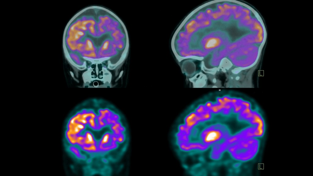 FDG uptake on PET scans
FDG uptake essentially describes the quantity of radiotracer that has been “taken up” or absorbed by cells in different parts of your body. Because different types of cells have varying metabolic demands, FDG will accumulate in different concentrations across the body. Areas with higher metabolic activity, meaning cells are working harder and using more glucose, will show a higher concentration of FDG.
FDG activity is closely related to FDG uptake and reflects the intensity with which tissues are utilizing glucose. It’s essentially a measure of the metabolic processes driving the absorption of FDG. While FDG uptake is the measurement of FDG present in tissues, FDG activity describes the metabolic characteristics that lead to this absorption. In simpler terms, higher FDG uptake generally indicates higher metabolic activity.
Now that we’ve established a basic understanding of FDG and uptake, let’s delve into the various terms you might find in your PET report related to FDG uptake levels.
“No Uptake” on a PET Scan: Interpreting the Absence of FDG Absorption
“No uptake” signifies that FDG is not being absorbed by the cells in a specific area. Whether this is a positive or negative finding depends heavily on the type of tissue being examined and the clinical context. In some tissues, low metabolic activity is normal and expected, while in others, it could indicate an abnormality. Your doctor will consider the tissue type, your overall health status, and your baseline scans (if available) to interpret this result accurately. Here are some potential interpretations of “no uptake”:
- Inactive Tumor or Growth: In areas previously known to have tumors or growths, “no uptake” could suggest that these abnormal tissues are no longer as metabolically active. This might indicate necrosis (tissue death) or, encouragingly, that treatments have been successful in reducing the growth’s activity. Further tests are usually needed to confirm these findings.
- Normal Tissue with Low Metabolic Activity: Certain tissues in the body naturally exhibit low metabolic activity and are expected to show little to no FDG uptake. In these cases, “no uptake” is a healthy and normal result.
- Technical Issues: In rare instances, “no uptake” could be due to technical limitations of the scanning equipment, such as insufficient sensitivity or malfunction.
- Resolved Inflammation or Infection: If the PET scan was performed to assess an area suspected of infection or inflammation, “no uptake” might suggest that the issue has resolved or is currently inactive.
“Normal FDG Uptake”: What to Expect in Healthy Tissues
Certain organs and tissues in your body, such as the brain, liver, and spleen, are naturally more metabolically active and tend to exhibit higher FDG uptake. These organs have higher glucose demands to perform their functions. Most tissues have established medical baselines for expected FDG absorption, which vary based on the tissue type and location within the body. “Normal uptake” results indicate that your tissues are functioning within these expected metabolic parameters, suggesting healthy activity.
“Mild” or “Low FDG Uptake”: When Lower Absorption Requires Further Attention
“Mild” or “low FDG uptake,” sometimes also described as “low-level” or “low-grade” uptake, can be normal for tissues that are inherently less metabolically active or those with a higher fat content. However, in areas where higher metabolic activity is expected, low uptake may warrant closer scrutiny. Potential explanations for low FDG uptake include:
- Less Active or Non-Viable Tissue: This could indicate that previously active areas are now necrotic or less functional. It might also suggest that a tumor, inflammation, or disease process is becoming less active or inactive, which could be a positive sign of treatment effectiveness. Your doctor will likely order further tests to clarify the situation.
- Tumor Characteristics: Some types of tumors or other conditions might naturally exhibit lower FDG uptake.
- Technical Limitations: As with “no uptake,” technical issues with the scanning equipment could sometimes contribute to falsely low readings.
“Increased FDG Uptake”: Understanding Higher Than Normal Absorption
“Increased FDG uptake” or “Intense FDG uptake” on a PET scan signifies that cells in a particular area are absorbing more FDG radiotracer than the surrounding tissues. This heightened uptake typically appears as brighter or more intense spots on the PET scan images. Increased FDG uptake can be indicative of several conditions:
- Cancer: Cancer cells are characteristically more metabolically active than normal cells, leading to increased glucose consumption and, consequently, higher FDG uptake.
- Inflammation or Infection: Areas experiencing active inflammation or infection often show increased FDG uptake due to the heightened metabolic activity of immune cells responding to the issue.
- Tissue Healing: Tissues undergoing repair after surgery, injury, or radiation therapy may temporarily exhibit increased FDG uptake as part of the healing process.
- Benign Conditions: Certain non-cancerous growths or active thyroid nodules can also demonstrate higher FDG uptake.
While increased FDG uptake is often a point of concern regarding potential cancer, it’s important to remember that it can also be caused by benign or non-malignant processes. Accurate interpretation by a radiologist, considering your clinical history and potentially ordering additional tests, is crucial for a correct diagnosis.
“Abnormal FDG Uptake”: Deviation from Expected Metabolic Activity
“Abnormal FDG uptake” indicates that glucose absorption in a tissue is at an irregular level compared to what is expected for that specific tissue type. This can mean either less than expected (lower than normal) or higher than expected (increased) FDG absorption. In cases of higher FDG uptake, further investigations are usually conducted to determine the underlying cause. Potential reasons for higher than expected “abnormal” FDG uptake include:
- Possible Cancer: Many types of cancer cells have elevated metabolic activity, resulting in a higher FDG absorption rate.
- Infection or Inflammation: As mentioned earlier, the increased metabolic activity of immune cells responding to infection or inflammation can also lead to higher FDG uptake.
SUV: Quantifying Uptake with the Standardized Uptake Value
SUV is a medical abbreviation for Standardized Uptake Value. It is a quantitative measure used in PET scans to determine the level of radiotracer activity in a specific area of the body at a particular time point. SUV is essentially a ratio that helps standardize the measurement of uptake, making it easier to compare scans performed at different times or on different machines. It is also referred to as the dose uptake ratio.
A higher SUV generally suggests increased metabolic activity, which, as we’ve discussed, could be due to various factors like inflammation, infection, or cancerous growths. Conversely, a lower SUV may indicate reduced metabolic activity. General ranges for SUV interpretation include:
- “Low intensity”: SUV less than 5
- “Moderate”: SUV between 5 and 10
- “Intense”: SUV between 10 and 15
- “Very intense”: SUV greater than 15
The SUV value is particularly valuable for monitoring disease progression or treatment response over time. Comparing SUV values from serial PET scans can provide radiologists with a clear understanding of how a condition or treatment is evolving.
Physiological Uptake: Normal Radiotracer Absorption in Healthy Organs
The term “physiological uptake” can sometimes be confusing because it is conceptually related to FDG uptake in general. The key distinction is that physiological uptake refers to the normal and expected absorption of any radiotracer throughout the body in healthy tissues. While PET scans often use FDG, physiological uptake is a broader concept applicable to any radiotracer. Different organs and tissues naturally have different standardized levels of radiotracer uptake because their metabolic activity varies by type and function.
Understanding these standard benchmarks for physiological uptake allows physicians to differentiate between normal organ function and potentially abnormal tissue behavior. Deviations from expected physiological uptake patterns may suggest the presence of disease, growths, or other health issues.
Physiological Uptake vs. Cancer: Distinguishing Normal from Abnormal
It’s crucial to understand that physiological uptake does not mean cancer. Physiological uptake is a normal phenomenon reflecting the expected absorption of a substance, like a radiotracer, by healthy tissues during imaging studies such as PET scans. This uptake naturally occurs in organs that actively use or process the radiotracer, such as the brain, heart, liver, and kidneys.
While increased uptake in certain areas can sometimes be a sign of cancer or other abnormalities, physiological uptake is considered a normal finding and is not indicative of disease. Radiologists and nuclear medicine specialists are trained to differentiate between physiological uptake and abnormal uptake when interpreting imaging results.
Physiological Activity in the Liver: Expected Liver Function on PET Scans
“Physiologic activity in the liver” refers to the normal metabolic activity occurring within the liver. This is an expected finding on a PET scan because the liver is a highly metabolically active organ with vital roles in glucose metabolism, detoxification, and protein synthesis. Here’s what “physiologic activity in the liver” generally means:
- Normal Finding: The liver naturally absorbs the FDG radiotracer due to its high metabolic rate. This is typically seen as a uniform or mildly increased uptake on the scan.
- Non-Specific: Physiological activity in the liver does not indicate any disease or pathology. It simply reflects the liver’s normal, healthy functioning.
- Normal Variations: The level of physiological uptake in the liver can vary slightly depending on factors like diet, blood sugar levels, and overall liver health, but it generally remains within a normal range.
Therefore, seeing “physiologic activity in the liver” on your PET scan report is a standard and reassuring finding, not indicative of any liver disease.
Physiological Activity in Kidneys and Bladder: Normal Excretion of Radiotracer
“Physiologic uptake in the kidneys and bladder” refers to the normal absorption and accumulation of the radiotracer in these organs during a PET scan. This is directly related to the body’s natural process of eliminating waste products.
- Kidneys: After the radiotracer is injected, it’s filtered out of the bloodstream by the kidneys as part of their waste removal function. This filtration process results in the normal, or physiologic, uptake of the radiotracer in the kidneys, which is visible on the PET scan.
- Bladder: As the kidneys filter the radiotracer, it’s excreted into the urine and subsequently accumulates in the bladder. The bladder then shows physiologic uptake as it stores this radiotracer before it is eliminated from the body through urination.
This physiologic uptake in the kidneys and bladder is expected and indicates that these organs are functioning correctly in filtering and excreting the radiotracer. It is not a sign of disease but rather a normal part of how the body processes and eliminates the radiotracer used in the PET scan.
Metabolic Activity on a PET Scan: Gauging Cellular Function
Metabolic activity on a PET scan refers to how actively cells in the body are using glucose or other metabolic substrates. PET scans detect this activity through the radiotracer, such as FDG, which highlights areas with increased metabolic uptake. This allows doctors to identify tissues that are more active than normal, which can be a sign of abnormal or diseased processes.
“No Metabolic Activity”: Interpreting the Absence of Cellular Function
“No metabolic activity” on a PET scan can have different implications depending on the location and context. In certain areas, it might be a normal finding, indicating tissues that naturally have low metabolic rates or don’t react significantly to glucose. However, if “no metabolic activity” is observed in an area where metabolic function is expected, it could suggest the following:
- Damaged or Necrotic Tissue: The tissue might be damaged or necrotic (dead) to the point where it is no longer metabolizing FDG effectively.
- Reduced Blood Flow or Blockage: Conditions that reduce blood flow to an area, such as heart attacks or strokes, can lead to a lack of metabolic activity.
- Successful Treatment Outcome: In the context of monitoring a tumor or growth, “no metabolic activity” can be a positive sign, suggesting that the abnormality is no longer active and that treatment has been successful.
“Low-Grade” or “Mild Metabolic Activity”: Reduced Cellular Function
“Low-grade metabolic activity,” also termed “mild metabolic activity,” indicates that less radiotracer is being absorbed than expected. For certain tissues, this can be a normal finding due to their inherent lower metabolic rate. However, when observed as an abnormality, low-grade metabolic activity can be associated with several conditions:
- Early Stages of Infections or Diseases: It might represent the initial phase of conditions like tumors or infections where metabolic activity hasn’t yet reached high levels.
- Scar Tissue: Scar tissue from previous surgeries or injuries typically has lower metabolic demands compared to healthy or diseased tissues.
- Aging Effects: Metabolic rate naturally tends to decline with age, so low-grade metabolic activity in older adults can sometimes be a normal age-related finding.
- Metabolic Disorders: Certain metabolic disorders, such as diabetes, metabolic syndrome, enzyme deficiencies, and hypothyroidism, can impact metabolic activity and radiotracer uptake.
- Early Response to Treatment: A reduction in metabolic activity might indicate that a tumor, inflammation, or disease is responding to treatment and beginning to heal.
“Increased Metabolic Activity” or “Hypermetabolic”: Elevated Cellular Function
“Increased metabolic activity,” also known as “hypermetabolic,” often suggests that cells are more active than normal and are consuming more glucose. This is frequently a sign of inflammation, infection, or cancer, as these conditions are often associated with increased cellular activity and glucose utilization.
“Increased metabolic activity” or “hypermetabolic” on a PET scan means that cells in a specific area are more active than usual, leading to increased radiotracer uptake and appearing as brighter spots on the scan images. This can indicate:
- Cancerous Tumors: Rapidly dividing cancer cells typically have high glucose demands.
- Inflammation and Infection: Active immune responses during inflammation or infection increase cellular activity.
- Healing and Tissue Repair: Regenerating tissues after injury or surgery can show temporary increases in metabolic activity.
- Benign Conditions: Some non-cancerous conditions, including benign tumors, may also exhibit hypermetabolic activity.
While increased metabolic activity can be a cause for concern, it is not always indicative of cancer. A radiologist and your doctor will interpret these results in the context of your medical history and potentially order further investigations to determine the cause.
Deauville Score: A Standard for Lymphoma Treatment Response
The Deauville score or scale (DS) is an internationally recognized standard used to report FDG uptake specifically in treatment trials for Hodgkin’s and non-Hodgkin’s lymphomas. Similar to SUV, it measures FDG uptake, but the Deauville score is a visual interpretation that compares uptake in affected areas to the physiological uptake in the liver and mediastinum (the space in the chest between the lungs).
The Deauville score ranges from 1 to 5:
- Score 1: No uptake in the area of concern.
- Score 2: Slight uptake, equal to or less than uptake in the mediastinum.
- Score 3: Uptake greater than the mediastinum but less than the liver.
- Score 4: Uptake slightly to moderately greater than the liver.
- Score 5: Markedly increased uptake compared to the liver.
In the Deauville scoring system, lower scores are better, indicating a better response to treatment. Scores 1 and 2 are generally considered complete responses, score 3 is adequate, while scores 4 and 5 are considered inadequate responses, suggesting the need for further treatment adjustments.
“Unremarkable” PET Scan Results: A Positive Finding
In medical terminology, “unremarkable” is generally a positive term. When your PET scan report states “unremarkable,” it means that the scan did not reveal any abnormal findings. In the context of a PET scan, being “unremarkable” is exactly what you hope for, indicating no signs of significant disease or abnormality detected by the scan.
PocketHealth Report Reader: Empowering You to Understand Your PET Scan
PocketHealth provides patients with fast and easy access to view, securely share, and store their PET scan results, often before a follow-up appointment with their physician.
If you encounter unfamiliar medical terms in your PET scan report, PocketHealth’s Report Reader tool offers definitions to help simplify complex medical jargon and make your report easier to understand.
As one PocketHealth user, Amy J., shared: “With Report Reader, I get explanations for words [in my report] that I don’t understand. I can now discuss my health with my family doctor with more confidence and have a better understanding of what is going on.”
The Value of Understanding Your PET Scan Results
PET scans are invaluable tools for visualizing the inner workings of your body and assessing organ function, playing a crucial role in diagnosing and monitoring various diseases. For patients like Jeanne, a breast cancer survivor, PocketHealth has been a lifeline, particularly when seeking medical care abroad. Having ready access to her imaging history enabled her to receive prompt and effective treatment for hip, back, and knee pain from a new doctor while in Costa Rica. This seamless access to her medical records ensured she received the necessary care, even when far from home.
The more you understand about your body and your health information, including your PET scan results, the more meaningful and productive your conversations with your healthcare team can be. Access to your PET scan results empowers you to ask more informed questions and participate more actively in decisions about your health journey. PocketHealth is committed to making this access easy and secure, helping you feel confident and in control every step of the way.