When your healthcare provider suggests a PET scan, understanding the results can feel like navigating a maze of medical terms. PET scan reports, filled with acronyms and technical language, might seem daunting. This guide aims to clarify common terms, including “physiologic uptake,” to help you better comprehend your report and engage in more informed discussions with your doctor.
While this information is intended to be helpful, it’s crucial to remember that it doesn’t replace your doctor’s professional interpretation. Your healthcare provider will discuss your results with you, either during a follow-up appointment or over the phone. However, familiarizing yourself with the terminology beforehand can empower you to ask more relevant questions and actively participate in your healthcare journey.
What is a PET Scan?
PET stands for Positron Emission Tomography. PET scans are a type of nuclear medicine imaging that utilizes a radioactive substance, known as a radiotracer, to create images of your body’s organs and tissues. During the procedure, a small amount of radiotracer is introduced into your body, either by injection, inhalation, or ingestion. This tracer emits positrons, which are detected by specialized cameras as you move through the PET scanner, a machine often described as donut-shaped. These recordings are then processed into detailed, color-coded images that a radiologist interprets.
PET scans are frequently used in conjunction with other imaging techniques like CT scans or MRIs. This combination allows for a comprehensive assessment of both the function and structure of organs and tissues, providing a more complete picture of your health.
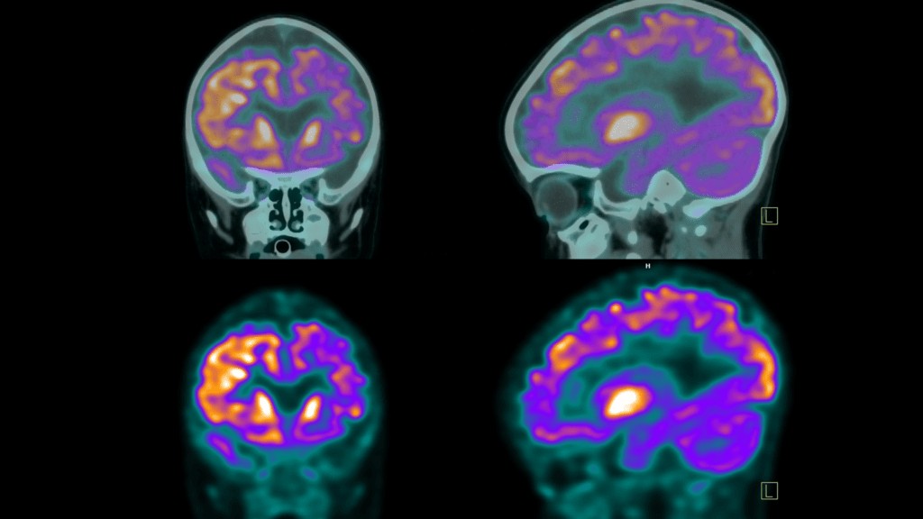
What are Axial, Sagittal, and Coronal Planes?
PET scanners capture images in three distinct planes, offering radiologists a 360-degree view of your internal anatomy:
- Axial Plane: Also known as the transverse view, this plane provides a horizontal cross-section, showing images of the body’s top and bottom portions. Imagine looking down at your body from above.
- Sagittal Plane: This is the side view, creating lateral images that divide the body into left and right halves.
- Coronal Plane: The frontal view, which shows images of the body’s front and back sections, as if you were facing someone directly.
Understanding these planes is crucial for visualizing the location of any findings described in your PET scan report.
What is a Radiotracer?
A radiotracer is a radioactive substance used in PET scans to highlight metabolic activity within the body. Often, a glucose-based radiotracer is used because many cells, especially those that are highly active, utilize glucose for energy. The most common radiotracer is Fluorodeoxyglucose (FDG).
The radiotracer is administered to the patient and then travels through the body. It accumulates in cells and tissues that are metabolically active, meaning they are using more energy. Your PET scan report will specify which radiotracer was used, the amount administered, and the method of administration (injection, inhalation, or ingestion).
What do FDG and FDG Uptake Mean on a PET Scan?
FDG stands for Fluorodeoxyglucose. It is a widely used radiotracer in PET scans because it mimics glucose, the body’s primary source of energy. Understanding FDG and the concept of “FDG uptake” is essential for interpreting your PET scan results.
“FDG uptake” or “FDG activity” refers to how cells absorb and utilize this radiotracer. Since different types of cells have varying metabolic rates, FDG will concentrate in different amounts in different areas of the body. Areas with higher metabolic activity, such as cancer cells or areas of inflammation, tend to absorb more FDG.
What is FDG Uptake?
 FDG uptake on PET scans
PET scan illustrating FDG uptake in the brain
FDG uptake quantifies the amount of radiotracer absorbed by cells. Different cell types have different energy requirements, leading to varying concentrations of FDG in different body regions. FDG activity reflects the intensity with which tissues are using glucose, indicated by the amount of FDG absorbed. Higher FDG activity usually signifies higher metabolic activity. Simply put, FDG uptake is a measure of FDG concentration in tissues, while FDG activity describes the metabolic processes causing FDG absorption.
Understanding FDG uptake is crucial for interpreting terms in your PET report. Let’s explore different levels of uptake and their potential meanings.
What does No Uptake Mean?
“No uptake” signifies that FDG is not being absorbed in a particular area. The interpretation of this finding depends on the specific tissue being examined and the clinical context.
- Inactive Tumor or Growth: In areas previously known to have tumors or growths, no uptake could indicate that the growth is no longer metabolically active. This might suggest successful treatment or tissue necrosis (cell death). Further tests are usually needed for confirmation.
- Normal Tissue: Some tissues naturally exhibit low metabolic activity and are expected to show little to no FDG uptake. In these cases, it is a normal and healthy finding.
- Technical Issues: In rare cases, “no uptake” could be due to technical problems with the scanning equipment or insufficient sensitivity.
- Resolved Inflammation/Infection: If the PET scan was intended to assess inflammation or infection, “no uptake” might suggest that the issue has resolved or is currently inactive.
What does Normal FDG Uptake Mean?
Certain organs and tissues, such as the brain, liver, and spleen, naturally exhibit higher FDG uptake due to their high glucose demands and metabolic activity. For most tissues, there are established medical baselines for expected FDG absorption, which vary depending on the tissue type and location. “Normal uptake” indicates that your tissues are functioning within these expected metabolic ranges.
What does Mild or Low FDG Uptake Mean?
“Low” or “mild” FDG uptake, sometimes referred to as “low-level” or “low-grade” uptake, can be normal for tissues with inherently lower metabolic activity, such as fatty tissues. However, in areas expected to be more active, it may warrant closer investigation.
- Less Active or Non-Viable Tissue: It could indicate that previously active tissues are now necrotic or that a tumor, inflammation, or disease is less active, possibly due to successful treatment. Further tests are usually needed for clarification.
- Certain Tumor Types: Some tumors naturally exhibit lower FDG uptake.
- Technical Limitations: As with “no uptake,” technical issues with the scanning equipment could also contribute to low uptake readings.
What does Increased FDG Uptake Mean?
“Increased FDG uptake” or “Intense FDG uptake” means cells in a specific area are absorbing more FDG radiotracer than the surrounding tissues. This appears as brighter or more intense spots on the PET scan images. Increased FDG uptake can be indicative of:
- Cancer: Cancer cells are typically highly metabolic and consume more glucose, resulting in increased FDG uptake.
- Inflammation or Infection: Areas of active inflammation or infection show increased uptake due to the heightened activity of immune cells.
- Tissue Healing: Tissues undergoing repair after surgery, injury, or radiation therapy may exhibit increased FDG uptake.
- Benign Conditions: Certain non-cancerous conditions, such as benign tumors or active thyroid nodules, can also show increased FDG uptake.
While increased FDG uptake can be concerning for cancer, it’s important to remember that it can also be caused by benign conditions. Accurate interpretation by a radiologist, considering your clinical history and other diagnostic tests, is crucial for proper diagnosis.
What does Abnormal FDG Uptake Mean?
“Abnormal FDG uptake” indicates that glucose absorption is outside the expected range for the tissue being assessed. This can mean either lower or higher than expected uptake. When higher FDG uptake is observed, further investigation is usually necessary to determine the cause. Possible reasons for higher abnormal FDG uptake include:
- Possible Cancer: Many cancer types exhibit increased metabolic activity and therefore higher FDG absorption.
- Infection or Inflammation: The increased metabolic activity of immune cells responding to infection or inflammation can also lead to higher FDG uptake.
What is SUV?
SUV is an abbreviation for Standardized Uptake Value. It is a quantitative measure used in PET scans to assess the level of radiotracer uptake in specific tissues. SUV is a ratio that compares the radiotracer activity in a region of interest to the radiotracer activity in the whole body, adjusted for body weight and injected dose. It is also referred to as the dose uptake ratio.
A higher SUV generally indicates increased metabolic activity, which could be due to various factors like inflammation, infection, or cancerous growths. Conversely, a lower SUV might suggest reduced metabolic activity. General SUV ranges for interpretation are:
- Low intensity: <5 SUV
- Moderate: 5-10 SUV
- Intense: 10-15 SUV
- Very intense: >15 SUV
SUV values are particularly helpful for comparing PET scans over time. Changes in SUV can provide valuable information about disease progression or treatment response.
What does Physiologic Uptake Mean on a PET Scan?
“Physiologic uptake” refers to the normal, expected absorption of a radiotracer throughout the body. It’s conceptually similar to FDG uptake but emphasizes the expected uptake in healthy tissues. While PET scans commonly use FDG, physiologic uptake isn’t specific to FDG; it applies to any radiotracer. The key concept is that different organs and tissues have established benchmarks for radiotracer uptake based on their inherent metabolic activity.
Understanding physiologic uptake benchmarks allows physicians to differentiate between normal organ function and potentially abnormal processes. Deviations from expected physiologic uptake patterns may indicate disease, growths, or other health issues. Therefore, when a report mentions “physiologic uptake,” it’s often reassuring, indicating normal function in those areas.
Does Physiologic Uptake Mean Cancer?
No, physiologic uptake does not mean cancer. It signifies normal radiotracer accumulation in tissues that naturally utilize or process the substance. Organs like the brain, heart, liver, and kidneys exhibit physiologic uptake because of their normal metabolic functions.
While increased uptake in certain areas can sometimes be associated with cancer, physiologic uptake is a normal finding and not indicative of disease. Distinguishing between physiologic and abnormal uptake is a critical part of PET scan interpretation, a task performed by radiologists and nuclear medicine specialists.
What does Physiological Activity in Liver Mean?
“Physiologic activity in the liver” describes normal metabolic function within the liver. The liver is a highly metabolically active organ responsible for numerous vital functions, including glucose metabolism, detoxification, and protein synthesis. Therefore, physiologic activity in the liver on a PET scan is expected and normal.
- Normal Finding: The liver naturally absorbs the radiotracer (like FDG) due to its high metabolic rate. This typically appears as uniform or mildly increased uptake on the scan.
- Non-Specific: Physiologic activity in the liver does not indicate any disease or abnormality. It simply reflects the liver’s normal function.
- Normal Variations: The level of uptake can vary slightly depending on factors such as diet, blood sugar levels, and overall liver health, but it generally remains within a normal range.
Physiologic liver activity on a PET scan is a standard, healthy finding.
What does Physiological Activity in Kidneys and Bladder Mean?
Physiologic uptake in the kidneys and bladder reflects the normal processing and excretion of the radiotracer.
- Kidneys: The kidneys filter the radiotracer from the bloodstream as part of the body’s waste removal process. This filtration results in normal radiotracer uptake in the kidneys, visible on the PET scan.
- Bladder: The filtered radiotracer is excreted into the urine and accumulates in the bladder. The bladder, therefore, shows physiologic uptake as it stores the radiotracer before elimination from the body.
This physiologic uptake in the kidneys and bladder is expected and indicates these organs are functioning correctly in filtering and excreting the radiotracer. It is not a sign of disease but rather a normal part of the PET scan process.
What does Metabolic Activity Mean on a PET Scan?
“Metabolic activity” on a PET scan refers to how actively cells in the body are using glucose or other metabolic substrates. PET scans detect this activity using a radiotracer like FDG, highlighting areas with increased metabolic uptake, which can help identify abnormal or diseased tissue. Physiologic uptake is a specific type of metabolic activity – the expected, normal metabolic activity of healthy organs.
What does No Metabolic Activity Mean on a PET Scan?
“No metabolic activity” can have different interpretations depending on the context. In some areas, it’s normal, but in others, it can be abnormal.
- Damaged Tissue: Tissue may be necrotic or damaged to the point where it no longer metabolizes FDG effectively.
- Blockages: Reduced blood flow or blockages, such as from heart attacks or strokes, can affect metabolic activity in an area.
- Successful Treatment: In the context of tumors, a lack of metabolic activity could indicate a positive treatment response and that the tumor is no longer active.
What does Low-Grade or Mild Metabolic Activity Mean?
“Low-grade metabolic activity” (or mild metabolic activity) indicates reduced radiotracer absorption. This can be normal for certain tissues, but when abnormal, it may suggest:
- Early Stages of Conditions: It could represent early-stage infections or diseases, including tumors, where metabolic activity is still low.
- Scar Tissue: Scar tissue from surgeries or injuries typically has lower metabolic demands.
- Aging: Metabolic rate naturally decreases with age, making low-grade activity normal in older adults.
- Metabolic Disorders: Conditions like diabetes or hypothyroidism can impact metabolic activity.
- Early Treatment Response: Reduced metabolic activity may indicate a positive response to treatment for tumors or inflammation.
What does Increased Metabolic Activity or Hypermetabolic Mean?
“Increased metabolic activity,” also called hypermetabolic, suggests cells are more active than normal and are consuming more glucose. This is often a sign of inflammation, infection, or cancer.
Increased metabolic activity indicates areas with higher-than-normal cellular activity, showing up as brighter spots on the PET scan due to increased radiotracer uptake. It can be caused by:
- Cancerous Tumors: Rapidly dividing cancer cells consume more glucose.
- Inflammation and Infection: Active immune responses increase cellular activity.
- Healing and Tissue Repair: Regenerating tissues post-injury or surgery can show temporary increases.
- Benign Conditions: Some non-cancerous conditions may also be hypermetabolic.
While increased metabolic activity can be concerning, it’s not always cancer. Radiologists and doctors will interpret these findings in the context of your overall health and further testing.
What’s a Deauville Score?
The Deauville score (DS) is a standardized scoring system used internationally to report FDG uptake in PET scans for Hodgkin’s and non-Hodgkin’s lymphomas, particularly in treatment trials. Like SUV, it measures FDG uptake, but it’s a visual assessment comparing uptake in affected areas to uptake in the liver and mediastinum (the space in the chest between the lungs).
The Deauville score ranges from 1 to 5:
- No uptake
- Slight uptake, equal to or below mediastinum uptake
- Uptake above mediastinum but below liver uptake
- Uptake slightly or moderately above liver uptake
- Uptake noticeably increased compared to liver uptake
For the Deauville score, lower is better. Scores of 1 and 2 are generally considered complete responses to treatment, while 3 is adequate, and 4 and 5 are inadequate.
Is Unremarkable Good or Bad?
In medical terminology, “unremarkable” is actually a positive finding. When your PET scan report states “unremarkable,” it means no significant abnormalities were detected. In the context of medical reports, unremarkable is exactly what you want to see.
Understanding Your PET Scan Results
Understanding the terms in your PET scan report can significantly empower you in your healthcare journey. While this guide provides explanations of key concepts like physiologic uptake and metabolic activity, it’s vital to discuss your specific results with your healthcare provider. They can provide personalized interpretation based on your medical history and clinical context. Armed with a better understanding of your report, you can engage more confidently in discussions about your health and make informed decisions in collaboration with your medical team.
PET scans are powerful tools that offer valuable insights into the body’s function. By demystifying some of the common terminology, this guide aims to make these insights more accessible and understandable, helping you navigate your healthcare with greater confidence.