When your veterinarian recommends a Positron Emission Tomography (PET) scan for your beloved pet, it’s natural to feel a mix of concern and a desire to understand what the results will reveal. PET scans are powerful diagnostic tools, but the reports can be filled with unfamiliar medical terms and acronyms. One term that might appear, and cause some confusion, is “physiologic activity.” This guide is designed to clarify what physiologic activity means on a PET scan in the context of your pet’s health, helping you navigate the report and have more informed discussions with your vet.
While this information is valuable for pet owners, it’s crucial to remember that understanding these terms is not a substitute for professional veterinary interpretation. Your veterinarian is best equipped to analyze your pet’s specific PET scan results in light of their overall health and medical history. Most vets will thoroughly review the findings with you during a consultation. However, by familiarizing yourself with some common PET scan terminology, you can be better prepared for these discussions, ask pertinent questions, and feel more involved in your pet’s care.
Understanding PET Scans for Pets
PET scans, short for Positron Emission Tomography, are a type of nuclear medicine imaging. They utilize a radioactive tracer to create detailed images of your pet’s internal organs and tissues, focusing on metabolic activity at a cellular level. This is particularly useful in veterinary medicine for detecting and monitoring various conditions, including cancer, neurological disorders, and inflammatory diseases.
During a PET scan, a small amount of a radiotracer, often a glucose-based substance like Fluorodeoxyglucose (FDG), is administered to your pet, usually through injection. This radiotracer travels through the bloodstream and is absorbed by tissues based on their metabolic activity – how actively cells are using energy. Tissues with higher metabolic rates, such as rapidly dividing cancer cells or areas of inflammation, will accumulate more of the radiotracer.
The PET scanner then detects the positrons emitted by the radiotracer within your pet’s body. This data is processed by a computer to create detailed, color-coded images. These images highlight areas of different metabolic activity, allowing veterinarians to visualize and assess the function of organs and tissues in a way that other imaging techniques, like X-rays or standard CT scans, cannot. Often, PET scans are used in conjunction with CT or MRI scans to provide both functional and anatomical information, giving a more complete picture of your pet’s condition.
Navigating PET Scan Views: Axial, Sagittal, and Coronal Planes
Understanding PET Scan Planes: Axial, Sagittal, and Coronal Views for Comprehensive Pet Imaging
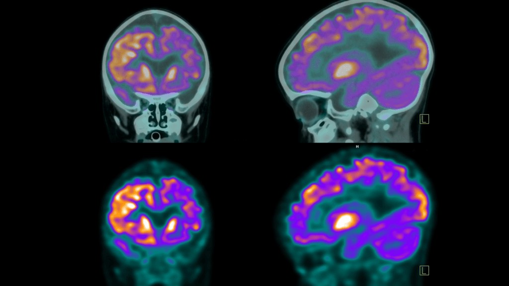
To provide a comprehensive view of your pet’s internal structures, PET scans capture images in three different planes:
- Axial Plane (Transverse View): Imagine looking down at your pet from above. The axial view provides horizontal cross-sections, showing images of the top and bottom portions of your pet’s body.
- Sagittal Plane (Lateral View): This is a side view, as if you were looking at your pet from the side. Sagittal images display the left and right halves of your pet’s body.
- Coronal Plane (Frontal View): Picture facing your pet head-on. The coronal view gives a face-on perspective, showing images of the front and back halves of your pet’s body.
These three perspectives are essential for radiologists and veterinarians to get a 360° understanding of your pet’s internal anatomy and identify any areas of interest from different angles.
Radiotracers in Pet PET Scans: The Key to Metabolic Insights
The radiotracer is a crucial component of a PET scan. It’s a specially designed substance, often a type of sugar attached to a radioactive atom, that allows metabolic activity to be visualized. In veterinary PET scans, as in human scans, Fluorodeoxyglucose (FDG) is the most commonly used radiotracer.
FDG is an analog of glucose, the body’s primary source of energy. Because cells that are highly metabolically active, such as cancer cells, inflammation sites, or active brain tissue, require more glucose, they will absorb more FDG. The radioactive component of FDG then emits positrons, which are detected by the PET scanner.
Your pet’s PET scan report will typically specify which radiotracer was used, the administered dose, the route of administration (injection, inhalation, or ingestion – though injection is most common in PET scans), and the location of administration. This information is important for accurate interpretation of the scan results.
FDG Uptake and Activity: Deciphering Your Pet’s PET Scan Report
Terms like “FDG uptake” and “FDG activity” are fundamental to understanding your pet’s PET scan report. They describe how cells absorb and utilize the FDG radiotracer, providing vital clues about metabolic processes within your pet’s body.
What is FDG Uptake in Pets?
 FDG uptake on PET scans
FDG uptake quantifies the amount of radiotracer absorbed by cells in specific tissues or organs. Different types of cells have varying metabolic demands. For instance, brain cells and active immune cells typically have higher glucose requirements compared to resting muscle tissue. Therefore, FDG will accumulate in different concentrations throughout your pet’s body.
FDG activity, closely related to uptake, refers to the intensity of glucose metabolism in tissues. Higher FDG activity generally indicates greater metabolic activity. In simpler terms, FDG uptake is the measured quantity of FDG in tissues, while FDG activity reflects the metabolic characteristics driving that absorption.
Understanding FDG uptake and activity is crucial for interpreting findings like “no uptake,” “normal uptake,” “mild uptake,” “increased uptake,” and importantly, “physiologic uptake,” which we will discuss in detail.
No Uptake: What Does it Mean for Your Pet?
“No uptake” or “absent uptake” signifies that FDG is not being absorbed in a particular area. The significance of this finding depends heavily on the tissue type being examined and the clinical context.
- Inactive Tumor or Growth: In areas previously known to have tumors or growths, no uptake can be a positive sign, suggesting that the tissue is no longer metabolically active. This could indicate successful treatment, necrosis (tissue death), or that the growth is dormant. However, further investigation is usually needed to confirm this.
- Normal Tissue: Some tissues naturally exhibit low metabolic activity. In these cases, no or minimal FDG uptake is considered normal and healthy.
- Technical Issues: Rarely, “no uptake” could be due to technical problems with the PET scanner’s sensitivity or malfunction.
- Resolved Inflammation/Infection: If a PET scan is performed to assess inflammation or infection, “no uptake” might suggest that the issue has resolved or is currently inactive.
Normal FDG Uptake in Pets: Expected Metabolic Activity
Certain organs and tissues in your pet’s body, such as the brain, liver, spleen, and kidneys, naturally exhibit higher FDG uptake due to their inherently higher metabolic rates. There are established medical baselines for expected FDG absorption in most tissues.
“Normal FDG uptake” indicates that the tissues are functioning within these expected metabolic parameters. It suggests healthy baseline activity for the specific organ or tissue being evaluated.
Mild or Low FDG Uptake: When is it Significant in Pets?
“Mild” or “low FDG uptake,” sometimes described as “low-level” or “low-grade,” can be normal for less metabolically active tissues, such as fat tissue or resting muscle. However, in areas expected to have higher metabolic activity, it might warrant closer attention.
- Less Active Tissue or Necrosis: Low uptake can indicate that previously active tissue is now necrotic or less viable. It could also signify that a tumor, inflammation, or disease process is regressing or becoming inactive, potentially due to successful treatment. Veterinarians will often order follow-up tests to clarify the situation.
- Certain Tumor Types: Some types of tumors naturally exhibit lower FDG avidity (tendency to absorb FDG).
- Technical Factors: Similar to “no uptake,” technical issues with the scanner could sometimes contribute to falsely low uptake readings.
Increased FDG Uptake: A Sign of Heightened Metabolic Activity in Pets
“Increased FDG uptake” or “intense FDG uptake” means that cells in a specific area are absorbing more radiotracer than the surrounding tissues. These areas appear brighter on the PET scan images. Increased uptake signifies heightened metabolic activity and can be associated with several conditions in pets:
- Cancer: Cancer cells are typically highly metabolic, consuming more glucose and thus exhibiting increased FDG uptake. This is a primary reason PET scans are valuable in cancer diagnosis and staging in veterinary oncology.
- Inflammation and Infection: Areas of active inflammation or infection demonstrate increased uptake due to the elevated metabolic activity of immune cells fighting the infection or driving the inflammatory process.
- Tissue Healing: Healing tissues, post-surgery, injury, or radiation therapy, can temporarily show increased FDG uptake as part of the repair process.
- Benign Conditions: Certain non-cancerous growths or active benign nodules can also exhibit increased FDG uptake.
While increased FDG uptake is often concerning for cancer, it’s crucial to understand that it’s not always indicative of malignancy. Benign conditions can also cause increased metabolic activity. Therefore, veterinary radiologists and your veterinarian will consider the clinical context, location of uptake, and other diagnostic findings to arrive at an accurate diagnosis.
Abnormal FDG Uptake: Deviation from Expected Metabolic Patterns
“Abnormal FDG uptake” indicates that glucose absorption is outside the expected range for the tissue being assessed. This can mean either lower or higher than anticipated uptake. When higher uptake is noted as abnormal, further investigation is usually required to determine the underlying cause, considering the possibilities mentioned above (cancer, infection, inflammation, etc.).
Standardized Uptake Value (SUV): Quantifying Metabolic Activity in Pets
SUV, or Standardized Uptake Value, is a quantitative measure used in PET scans to assess the degree of radiotracer uptake in a specific area of interest. It’s a ratio that standardizes the measurement of radiotracer activity, taking into account factors like the injected dose and your pet’s body weight.
A higher SUV generally indicates increased metabolic activity. While specific SUV thresholds can vary based on the type of PET scan and the clinical context, general ranges are often used for interpretation:
- Low Intensity: SUV < 5
- Moderate: SUV 5-10
- Intense: SUV 10-15
- Very Intense: SUV > 15
SUV values are particularly useful for comparing PET scans over time, such as monitoring treatment response. Changes in SUV can provide objective evidence of whether a condition is progressing, stable, or responding to therapy.
Physiologic Uptake: Normal Metabolic Activity on a PET Scan
Now, let’s focus on the key term: “physiologic uptake.” This term describes the expected, normal absorption of the radiotracer in various organs and tissues throughout your pet’s body. It’s crucial to understand that different organs naturally have different levels of metabolic activity.
Physiologic uptake reflects the baseline metabolic function of healthy tissues. For example, the brain, heart, liver, kidneys, and bladder are normally metabolically active and will therefore show physiologic uptake of FDG. This uptake is not indicative of disease; it simply represents the normal functioning of these organs.
Physiologic Uptake vs. Abnormal Uptake: Key Differences for Pet Owners
The critical distinction is between physiologic uptake and abnormal uptake. Physiologic uptake is expected and normal, while abnormal uptake suggests a deviation from the typical metabolic pattern and may indicate a problem.
Physiologic uptake is typically:
- Predictable: It occurs in organs known to be metabolically active.
- Symmetrical: Often seen bilaterally in paired organs like kidneys.
- Consistent: Generally within a known range for a given organ.
Abnormal uptake, in contrast, is:
- Unexpected: Occurs in areas where high metabolic activity is not normally seen.
- Asymmetrical: May be focal or localized to one area.
- Intense: Often significantly higher than background or physiologic uptake levels.
Does Physiologic Uptake Mean Cancer in Pets?
No, physiologic uptake does not mean cancer. It is a normal finding representing the healthy metabolic function of organs. It’s essential not to confuse physiologic uptake with abnormal or increased uptake, which may raise suspicion for cancer or other diseases.
Physiologic Activity in the Liver, Kidneys, and Bladder of Pets: Common Normal Findings
Understanding physiologic uptake in specific organs is helpful. Here’s what it means for the liver, kidneys, and bladder, organs commonly showing physiologic activity on PET scans:
Physiologic Activity in the Liver
Normal Liver Activity on PET Scan: Illustrating Physiologic Uptake
“Physiologic activity in the liver” is a normal finding. The liver is a highly metabolic organ responsible for numerous functions, including glucose metabolism, detoxification, and protein synthesis.
- Normal Finding: The liver naturally absorbs FDG due to its metabolic role. This appears as uniform or mildly increased uptake on the PET scan.
- Non-Specific: Physiologic activity in the liver does not indicate any disease or abnormality.
- Variations: The level of uptake can vary slightly based on factors like diet and overall liver health, but it remains within a normal physiologic range.
Physiologic Activity in Kidneys and Bladder
Kidney and Bladder Activity on PET Scan: Demonstrating Normal Physiologic Uptake
Physiologic uptake in the kidneys and bladder is also expected and related to the body’s excretion of the radiotracer.
- Kidneys: The kidneys filter the FDG radiotracer from the bloodstream as part of waste removal. This normal filtration process leads to physiologic uptake in the kidneys, visible on the PET scan.
- Bladder: As the kidneys filter FDG, it’s excreted into the urine and accumulates in the bladder. The bladder therefore shows physiologic uptake as it stores the radiotracer before elimination from the body.
This uptake in the kidneys and bladder is a sign that these organs are functioning correctly in processing and eliminating waste and the radiotracer. It is not indicative of disease.
Metabolic Activity: The Broader Context of PET Scan Interpretation for Pets
“Metabolic activity” is the overarching concept that PET scans are designed to assess. It refers to how actively cells use glucose or other metabolic substrates to function. By detecting radiotracer uptake, PET scans visualize and measure this activity, helping identify areas of abnormal or potentially diseased tissue in your pet.
No Metabolic Activity: Implications for Your Pet’s Health
“No metabolic activity,” when noted as an abnormality, can suggest several possibilities:
- Damaged or Necrotic Tissue: Tissue damage or necrosis can lead to a lack of metabolic activity in an area that would normally be active.
- Reduced Blood Flow: Blockages or reduced blood supply, such as from a stroke, can decrease metabolic activity in the affected region.
- Successful Treatment: In the context of tumors, no metabolic activity can be a positive sign of successful treatment, indicating the tumor is no longer active.
Low-Grade or Mild Metabolic Activity: Subtle Changes in Pet PET Scans
“Low-grade” or “mild metabolic activity” indicates reduced radiotracer uptake. While normal in some tissues, as an abnormal finding, it might suggest:
- Early Stages of Disease: Early infections or tumors may exhibit low-grade activity before metabolic activity increases significantly.
- Scar Tissue: Scar tissue has lower metabolic demands compared to healthy or diseased tissue.
- Age-Related Changes: Metabolic rate can naturally decline with age.
- Metabolic Disorders: Certain metabolic disorders can affect overall metabolic activity.
- Treatment Response: Decreasing metabolic activity can indicate a positive response to treatment.
Increased Metabolic Activity or Hypermetabolic: Areas of Concern in Pet PET Scans
“Increased metabolic activity” or “hypermetabolic” signifies that cells are more active than normal and are consuming more glucose. This is a significant finding that often raises concerns about:
- Cancerous Tumors: Rapidly dividing cancer cells are highly metabolic.
- Inflammation and Infection: Active immune responses drive increased cellular activity.
- Healing and Tissue Repair: Repair processes increase metabolic demands temporarily.
- Benign Conditions: Some non-cancerous conditions can also be hypermetabolic.
Increased metabolic activity is a key indicator of potential issues, but it requires careful interpretation in conjunction with other clinical information.
Deauville Score: Assessing Lymphoma Treatment Response in Pets
The Deauville score is a standardized scoring system, primarily used in lymphoma, to visually assess FDG uptake and treatment response. It compares FDG uptake in affected areas to normal background uptake in the liver and mediastinum. The Deauville scale ranges from 1 to 5, with lower scores indicating better treatment response. While originally developed for human lymphoma, it can be adapted and utilized in veterinary oncology for assessing lymphoma in pets.
“Unremarkable” PET Scan Results: Good News for Your Pet
In medical reports, “unremarkable” is generally positive. An “unremarkable” PET scan report means that no significant abnormalities were detected. This is the desired outcome – an unremarkable PET scan suggests no evidence of concerning metabolic activity.
Empowering Pet Owners with Knowledge
Understanding your pet’s PET scan results can feel empowering. While professional veterinary interpretation is essential, being familiar with terms like “physiologic activity,” “FDG uptake,” and “SUV” allows you to participate more actively in discussions about your pet’s health and care. By asking informed questions and understanding the nuances of the report, you can work collaboratively with your veterinarian to make the best decisions for your furry companion. Access to clear and understandable explanations of medical terms can significantly improve your experience and confidence in navigating your pet’s healthcare journey.