What Does Pet Measure? Positron Emission Tomography (PET) scans measure vital physiological functions, offering valuable insights into blood flow, metabolic activity, neurotransmitter interactions, and the behavior of radiolabeled drugs in pets. PET scans provide quantitative data, enabling the monitoring of changes during disease progression or in response to specific treatments. Discover how PET scans can revolutionize pet healthcare with detailed information available at PETS.EDU.VN, your reliable source for pet health and diagnostics. Learn about various applications, including tumor detection and neurological assessments.
1. Understanding Positron Emission Tomography (PET) in Veterinary Medicine
Positron Emission Tomography (PET) scans are advanced imaging techniques that play a pivotal role in modern veterinary medicine. Unlike traditional imaging methods like X-rays or CT scans, which primarily visualize anatomical structures, PET scans delve into the functional aspects of a pet’s body. Specifically, what does PET measure? It assesses physiological functions by examining blood flow, metabolic rates, neurotransmitter activity, and the distribution of radiolabeled drugs. This capability provides a comprehensive understanding of how different body parts are working at a cellular level, which is crucial for diagnosing and managing various health conditions in pets.
PET scans offer several advantages over other imaging techniques. For instance, the quantitative nature of PET allows for the precise measurement of changes over time. This is especially valuable in monitoring the progression of diseases and evaluating the effectiveness of treatments. Moreover, PET scans can detect abnormalities at an early stage, often before structural changes are visible on X-rays or CT scans. This early detection can lead to more timely and effective interventions, significantly improving a pet’s prognosis.
1.1. The Science Behind PET Scans
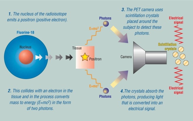
The science behind PET scans involves the detection of radioactivity emitted after a small amount of a radioactive tracer is injected into a peripheral vein of the pet. This tracer, typically labeled with isotopes such as oxygen-15, fluorine-18, carbon-11, or nitrogen-13, emits positrons as it decays. When a positron collides with an electron in the body, it produces two gamma rays that travel in opposite directions. These gamma rays are detected by the PET scanner, which then creates a detailed image of the tracer’s distribution within the body.
The tracer used in a PET scan is carefully selected based on the specific physiological process being investigated. For example, 18-fluorodeoxyglucose (FDG), a glucose analog, is commonly used to measure glucose metabolism. Since malignant tumors often metabolize glucose at a faster rate than benign tissues, FDG-PET scans are particularly useful in cancer diagnosis and staging. Similarly, different tracers can be used to assess blood flow in the brain, neurotransmitter activity, and other vital functions.
The total radioactive dose administered during a PET scan is similar to that used in computed tomography (CT) scans. While radiation exposure is always a concern, the benefits of PET scans in terms of diagnosis and treatment planning generally outweigh the risks. Veterinarians and radiologists carefully consider the potential risks and benefits before recommending a PET scan for a pet.
1.2. Applications of PET Scans in Veterinary Medicine
PET scans have a wide range of applications in veterinary medicine, making them an indispensable tool for diagnosing and managing various conditions. Some of the most common applications include:
- Oncology: PET scans are frequently used to diagnose, stage, and monitor cancer in pets. The ability to measure glucose metabolism allows for the detection of tumors, assessment of their aggressiveness, and evaluation of treatment response. PET scans can also help differentiate between recurrent tumors and post-treatment changes like radiation necrosis or surgical scarring.
- Neurology: PET scans can provide valuable information about blood flow, oxygen consumption, and neurotransmitter activity in the brain. This is particularly useful in understanding conditions such as strokes, dementia, and Parkinson’s disease in animals. PET scans can also help identify areas of abnormal brain activity that may be associated with seizures or behavioral changes.
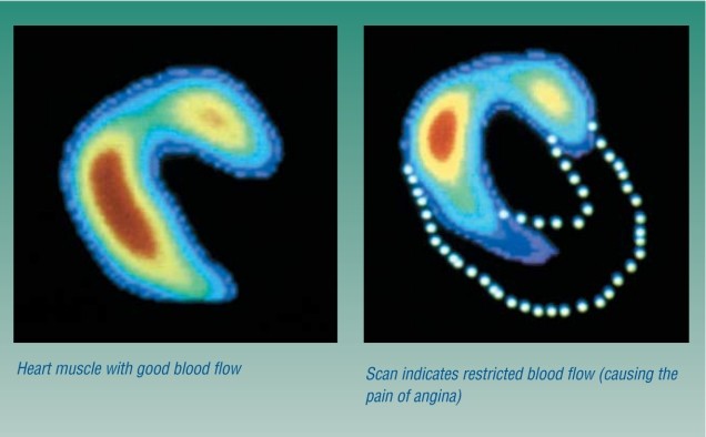
- Cardiology: In cardiology, PET scans can be used to assess the viability of heart muscle tissue, particularly in pets with heart disease. This information can help guide treatment decisions, such as whether a pet is a candidate for a heart transplant or other interventions.
- Inflammation and Infection: PET scans can detect areas of inflammation and infection throughout the body. This can be helpful in diagnosing conditions such as inflammatory bowel disease, arthritis, and infections that are difficult to detect with other imaging methods.
1.3. Benefits of PET Scans for Pets
The benefits of PET scans for pets are numerous. One of the most significant advantages is the ability to detect diseases at an early stage, often before clinical signs are evident. This early detection can lead to more effective treatment and improved outcomes for pets. Additionally, PET scans can provide valuable information about the extent and severity of a disease, helping veterinarians tailor treatment plans to the individual needs of each pet.
PET scans are also non-invasive, meaning they do not require surgery or other invasive procedures. This reduces the risk of complications and makes the procedure more comfortable for pets. The scans typically take between 10 and 40 minutes to complete, and pets remain fully clothed during the process. While some pets may experience mild anxiety during the scan, sedation is generally not required.
Overall, PET scans are a valuable tool in veterinary medicine, providing critical information that can improve the health and well-being of pets. At PETS.EDU.VN, we are committed to providing pet owners with the latest information about PET scans and other advanced diagnostic techniques. Our team of experts can help you understand the benefits of PET scans and determine whether this procedure is right for your pet. For more information, please visit our website or contact us at 789 Paw Lane, Petville, CA 91234, United States, or call us at +1 555-987-6543.
2. The PET Scan Procedure: A Step-by-Step Guide for Pet Owners
Understanding the PET scan procedure can help alleviate any anxiety you might have about your pet undergoing this diagnostic test. PET scans are non-invasive and generally well-tolerated by animals. Here’s a detailed, step-by-step guide to what you can expect during the process.
2.1. Preparation Before the PET Scan
Prior to the PET scan, your veterinarian will provide specific instructions to prepare your pet. These instructions are crucial for ensuring the accuracy and success of the scan. Common preparations include:
- Fasting: Your pet may need to fast for several hours before the scan. This is because food can interfere with the distribution of the radioactive tracer, particularly when measuring glucose metabolism. The duration of the fast will depend on the type of tracer used and the specific goals of the scan. Typically, this ranges from 4 to 12 hours.
- Hydration: Ensuring your pet is well-hydrated is important for optimal image quality. Dehydration can affect blood flow and tracer distribution, leading to inaccurate results. Your veterinarian may recommend giving your pet extra water in the hours leading up to the scan.
- Medication: Discuss all medications your pet is currently taking with your veterinarian. Some medications can interfere with the PET scan, and your veterinarian may advise temporarily discontinuing them before the procedure. Do not stop any medication without consulting your veterinarian first.
- Comfort: Make sure your pet is comfortable and relaxed before the scan. A calm pet is more likely to cooperate during the procedure, reducing the need for sedation. Bring a favorite toy or blanket to help your pet feel more at ease.
2.2. The PET Scan Procedure: What to Expect
The PET scan procedure itself is relatively straightforward. Here’s what typically happens:
- Arrival and Check-In: Upon arrival at the veterinary facility, you will check in and complete any necessary paperwork. The veterinary staff will review the procedure with you and answer any questions you may have.
- Tracer Injection: A small amount of radioactive tracer is injected into a peripheral vein of your pet. The tracer is administered intravenously, usually in the leg or paw. The injection is quick and generally painless.
- Waiting Period: After the tracer is injected, there is a waiting period of approximately 30 to 60 minutes. This allows the tracer to distribute throughout your pet’s body and accumulate in the tissues of interest. During this time, your pet will need to remain still to ensure accurate imaging.
- Scanning: Once the waiting period is over, your pet will be positioned on the PET scanner bed. The scanner is a large, donut-shaped machine that surrounds your pet. The scanning process takes between 10 and 40 minutes, depending on the area being imaged and the specific goals of the scan.
- During the Scan: During the scan, it is crucial that your pet remains as still as possible. Movement can blur the images and reduce their accuracy. In some cases, sedation may be necessary to ensure your pet remains still. The veterinary staff will monitor your pet closely throughout the scan.
- Completion: Once the scan is complete, your pet can be taken home. There are usually no special precautions required after the scan. However, your veterinarian may advise you to monitor your pet for any unusual behavior or signs of discomfort.
2.3. After the PET Scan: Recovery and Results
After the PET scan, your pet can typically resume normal activities. The radioactive tracer used in the scan has a short half-life, meaning it decays quickly and is eliminated from the body within a few hours. Your veterinarian will provide specific instructions regarding post-scan care, if necessary.
The results of the PET scan will be reviewed by a radiologist, who will analyze the images and prepare a report for your veterinarian. The report will describe the distribution of the tracer in your pet’s body and identify any areas of abnormal activity. Your veterinarian will discuss the results with you and explain their implications for your pet’s health.
Depending on the findings of the PET scan, your veterinarian may recommend further diagnostic tests or treatments. The PET scan results can help guide treatment decisions and improve the overall management of your pet’s condition.
At PETS.EDU.VN, we understand that undergoing a PET scan can be a stressful experience for both you and your pet. Our goal is to provide you with the information and support you need to make informed decisions about your pet’s health. If you have any questions or concerns about the PET scan procedure, please do not hesitate to contact us. You can reach us at 789 Paw Lane, Petville, CA 91234, United States, or call us at +1 555-987-6543. Visit our website at PETS.EDU.VN for more information about PET scans and other diagnostic services.
3. Interpreting PET Scan Results: What They Mean for Your Pet
Interpreting PET scan results requires expertise and a thorough understanding of your pet’s medical history. Radiologists and veterinarians work together to analyze the images and determine the significance of any abnormalities detected. Here’s a guide to help you understand what PET scan results can reveal.
3.1. Understanding Tracer Uptake
The key to interpreting PET scan results lies in understanding tracer uptake. Different tissues and organs in the body absorb the radioactive tracer at different rates. The amount of tracer uptake reflects the level of metabolic activity in that area. Areas with high metabolic activity, such as tumors, tend to accumulate more tracer than areas with low metabolic activity.
The radiologist will examine the PET scan images to identify areas of increased or decreased tracer uptake. These areas are often referred to as “hot spots” (increased uptake) or “cold spots” (decreased uptake). The location, size, and intensity of these spots provide valuable information about the underlying condition.
For example, in cancer diagnosis, a “hot spot” on a PET scan may indicate the presence of a tumor. The radiologist will assess the size and location of the tumor, as well as its metabolic activity, to determine its aggressiveness and stage. This information is crucial for planning the most effective treatment strategy.
3.2. Common Findings and Their Implications
PET scans can reveal a variety of findings, each with its own implications for your pet’s health. Some of the most common findings include:
- Tumors: PET scans are highly sensitive for detecting tumors, even those that are small or difficult to locate with other imaging methods. The scan can also differentiate between benign and malignant tumors based on their metabolic activity. Malignant tumors typically exhibit higher glucose metabolism and therefore show greater tracer uptake.
- Inflammation: PET scans can detect areas of inflammation throughout the body. This can be helpful in diagnosing conditions such as inflammatory bowel disease, arthritis, and infections. The scan can also assess the severity and extent of the inflammation, which can guide treatment decisions.
- Infection: Similar to inflammation, PET scans can identify areas of infection in the body. This is particularly useful for detecting deep-seated infections that are difficult to diagnose with other methods. The scan can also help determine the extent of the infection and monitor the response to treatment.
- Neurological Disorders: PET scans can provide valuable information about brain function, including blood flow, oxygen consumption, and neurotransmitter activity. This can be helpful in diagnosing and managing neurological disorders such as strokes, dementia, and seizures.
- Cardiovascular Disease: In cardiology, PET scans can assess the viability of heart muscle tissue. This is particularly useful in pets with heart disease, as it can help determine whether a pet is a candidate for a heart transplant or other interventions.
3.3. Working with Your Veterinarian
The interpretation of PET scan results is a complex process that requires the expertise of both a radiologist and a veterinarian. The radiologist will analyze the images and prepare a report, while the veterinarian will integrate the findings with your pet’s medical history and clinical signs.
It is important to discuss the PET scan results with your veterinarian in detail. Ask questions about any abnormalities detected and their potential implications for your pet’s health. Your veterinarian will explain the findings in a way that you can understand and discuss the next steps in your pet’s care.
Depending on the PET scan results, your veterinarian may recommend further diagnostic tests, such as biopsies or blood tests. They may also recommend specific treatments, such as surgery, chemotherapy, or medication. The PET scan results can help guide these decisions and improve the overall management of your pet’s condition.
At PETS.EDU.VN, we are committed to providing pet owners with the information and support they need to understand PET scan results and make informed decisions about their pet’s health. Our team of experts can help you navigate the complexities of PET scan interpretation and ensure that your pet receives the best possible care. Contact us at 789 Paw Lane, Petville, CA 91234, United States, or call us at +1 555-987-6543. Visit our website at PETS.EDU.VN for more information about PET scans and other diagnostic services.
4. The Role of PET Scans in Cancer Diagnosis and Staging for Pets
PET scans have revolutionized cancer diagnosis and staging in both human and veterinary medicine. Their ability to detect metabolic activity at the cellular level makes them invaluable for identifying tumors, assessing their aggressiveness, and determining the extent of disease. Here’s how PET scans play a critical role in cancer care for pets.
4.1. Early Detection of Tumors
One of the most significant benefits of PET scans is their ability to detect tumors at an early stage, often before they are visible on other imaging methods. This early detection can lead to more timely and effective interventions, significantly improving a pet’s prognosis.
PET scans use radioactive tracers, such as 18-fluorodeoxyglucose (FDG), to measure glucose metabolism. Cancer cells typically metabolize glucose at a faster rate than normal cells, so they accumulate more FDG. This allows PET scans to identify tumors as “hot spots” of increased metabolic activity.
The sensitivity of PET scans makes them particularly useful for detecting small tumors or tumors in difficult-to-reach areas. They can also differentiate between benign and malignant tumors based on their metabolic activity. Malignant tumors typically exhibit higher glucose metabolism and therefore show greater FDG uptake.
4.2. Accurate Staging of Cancer
Accurate staging of cancer is essential for determining the most appropriate treatment strategy. Staging involves assessing the size and location of the primary tumor, as well as whether the cancer has spread to other parts of the body. PET scans can provide valuable information for staging cancer in pets.
PET scans can detect metastatic tumors, which are tumors that have spread from the primary site to distant organs or tissues. This is particularly important for cancers that tend to metastasize early, such as lymphoma and osteosarcoma. The scan can identify metastatic tumors that may not be visible on other imaging methods, such as X-rays or CT scans.
By providing a comprehensive assessment of the extent of disease, PET scans can help veterinarians determine the stage of cancer. This information is crucial for selecting the most effective treatment approach, whether it involves surgery, chemotherapy, radiation therapy, or a combination of these modalities.
4.3. Monitoring Treatment Response
PET scans are also valuable for monitoring treatment response in pets with cancer. The scan can assess whether a tumor is responding to treatment by measuring changes in its metabolic activity. A decrease in FDG uptake indicates that the tumor is becoming less metabolically active and is responding to treatment.
PET scans can also help differentiate between recurrent tumors and post-treatment changes, such as radiation necrosis or surgical scarring. This can be particularly challenging with other imaging methods, as these changes can look similar on X-rays or CT scans. PET scans can distinguish between these conditions based on their metabolic activity.
By monitoring treatment response with PET scans, veterinarians can adjust treatment plans as needed to optimize outcomes for pets with cancer. This can involve changing the dose or type of chemotherapy, adding radiation therapy, or considering other interventions.
At PETS.EDU.VN, we are dedicated to providing pet owners with the latest information about cancer diagnosis and treatment in pets. Our team of experts can help you understand the role of PET scans in cancer care and make informed decisions about your pet’s health. You can reach us at 789 Paw Lane, Petville, CA 91234, United States, or call us at +1 555-987-6543. Visit our website at PETS.EDU.VN for more information about PET scans and other diagnostic services.
5. PET Scans in Neurological Assessments for Pets
PET scans are increasingly utilized in veterinary neurology to assess brain function and diagnose neurological disorders in pets. By measuring blood flow, oxygen consumption, and neurotransmitter activity, PET scans can provide valuable insights into the underlying causes of neurological symptoms.
5.1. Assessing Brain Function
PET scans can assess various aspects of brain function, including:
- Blood Flow: PET scans can measure blood flow to different regions of the brain. Decreased blood flow may indicate a stroke, while increased blood flow may indicate inflammation or seizure activity.
- Oxygen Consumption: PET scans can measure the rate at which different regions of the brain consume oxygen. This can be helpful in identifying areas of tissue damage or dysfunction.
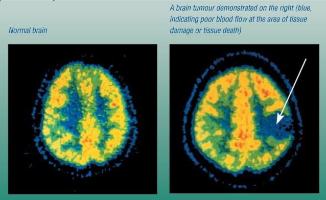
- Glucose Metabolism: As mentioned earlier, PET scans can measure glucose metabolism in the brain. This is particularly useful for detecting tumors, as cancer cells typically metabolize glucose at a faster rate than normal cells.
- Neurotransmitter Activity: PET scans can measure the activity of neurotransmitters, such as dopamine, serotonin, and norepinephrine. This can be helpful in diagnosing and managing neurological disorders such as Parkinson’s disease and depression.
By providing a comprehensive assessment of brain function, PET scans can help veterinarians diagnose neurological disorders and develop appropriate treatment plans.
5.2. Diagnosing Neurological Disorders
PET scans can be used to diagnose a variety of neurological disorders in pets, including:
- Strokes: PET scans can detect areas of decreased blood flow in the brain, which may indicate a stroke. The scan can also assess the extent of the damage and monitor the recovery process.
- Dementia: PET scans can detect changes in brain metabolism that may indicate dementia. The scan can also help differentiate between different types of dementia, such as Alzheimer’s disease and vascular dementia.
- Seizures: PET scans can identify areas of abnormal brain activity that may be associated with seizures. The scan can also help determine the type of seizure and guide treatment decisions.
- Brain Tumors: PET scans can detect brain tumors and assess their aggressiveness. The scan can also help differentiate between benign and malignant tumors and guide treatment planning.
- Parkinson’s Disease: PET scans can measure dopamine activity in the brain, which is reduced in Parkinson’s disease. The scan can help diagnose the disease and monitor the response to treatment.
5.3. Guiding Treatment Decisions
PET scans can help guide treatment decisions for pets with neurological disorders. The scan can provide valuable information about the underlying cause of the symptoms and the extent of the damage. This information can help veterinarians select the most appropriate treatment approach, whether it involves medication, surgery, or other interventions.
For example, in pets with seizures, PET scans can help identify the area of the brain where the seizures originate. This information can guide surgical removal of the affected tissue, which may be an option for pets with refractory seizures.
In pets with brain tumors, PET scans can help determine the type of tumor and its aggressiveness. This information can guide treatment planning, such as whether to pursue surgery, radiation therapy, or chemotherapy.
At PETS.EDU.VN, we are committed to providing pet owners with the latest information about neurological assessments in pets. Our team of experts can help you understand the role of PET scans in diagnosing and managing neurological disorders and make informed decisions about your pet’s health. Contact us at 789 Paw Lane, Petville, CA 91234, United States, or call us at +1 555-987-6543. Visit our website at PETS.EDU.VN for more information about PET scans and other diagnostic services.
6. PET Scans in Cardiology: Assessing Heart Health in Pets
PET scans are not just for cancer and neurological disorders; they also play a significant role in assessing heart health in pets. In cardiology, PET scans can be used to evaluate blood flow to the heart muscle, assess the viability of heart tissue, and diagnose various cardiovascular conditions.
6.1. Evaluating Blood Flow to the Heart Muscle
PET scans can measure blood flow to different regions of the heart muscle. This is particularly important in pets with coronary artery disease, where the arteries that supply blood to the heart become narrowed or blocked. Decreased blood flow to the heart muscle can lead to chest pain (angina), heart attack, and other complications.
PET scans use radioactive tracers, such as rubidium-82 or nitrogen-13 ammonia, to measure blood flow to the heart. The tracer is injected into the bloodstream, and the PET scanner detects the amount of tracer that reaches different regions of the heart muscle. Areas with decreased blood flow will show reduced tracer uptake.
6.2. Assessing Viability of Heart Tissue
In pets with heart disease, PET scans can assess the viability of heart tissue. This is particularly important in pets who have had a heart attack or have chronic heart failure. PET scans can help determine whether the heart muscle is still viable and can potentially recover with treatment.
PET scans use radioactive tracers, such as FDG, to measure glucose metabolism in the heart muscle. Viable heart tissue will metabolize glucose, while non-viable tissue will not. PET scans can differentiate between these two types of tissue and help guide treatment decisions.
For example, if a PET scan shows that a significant portion of the heart muscle is still viable, a veterinarian may recommend treatments such as angioplasty or bypass surgery to improve blood flow to the heart. If the heart muscle is not viable, these treatments may not be effective.
6.3. Diagnosing Cardiovascular Conditions
PET scans can be used to diagnose a variety of cardiovascular conditions in pets, including:
- Coronary Artery Disease: PET scans can detect decreased blood flow to the heart muscle, which may indicate coronary artery disease.
- Heart Failure: PET scans can assess the viability of heart tissue in pets with heart failure.
- Cardiomyopathy: PET scans can help diagnose different types of cardiomyopathy, which are diseases of the heart muscle.
- Myocardial Infarction (Heart Attack): PET scans can detect areas of damage in the heart muscle after a heart attack.
By providing a comprehensive assessment of heart health, PET scans can help veterinarians diagnose cardiovascular conditions and develop appropriate treatment plans for pets.
At PETS.EDU.VN, we are dedicated to providing pet owners with the latest information about cardiac assessments in pets. Our team of experts can help you understand the role of PET scans in diagnosing and managing cardiovascular conditions and make informed decisions about your pet’s health. Contact us at 789 Paw Lane, Petville, CA 91234, United States, or call us at +1 555-987-6543. Visit our website at PETS.EDU.VN for more information about PET scans and other diagnostic services.
7. Risks and Considerations of PET Scans for Pets
While PET scans offer numerous benefits in veterinary medicine, it’s essential to be aware of the potential risks and considerations associated with the procedure. Understanding these factors can help you make informed decisions about your pet’s health.
7.1. Radiation Exposure
PET scans involve the use of radioactive tracers, which expose your pet to a small amount of radiation. While the radiation dose is generally considered safe, it’s important to minimize exposure as much as possible.
The radiation dose from a PET scan is similar to that of a CT scan. The radioactive tracer has a short half-life, meaning it decays quickly and is eliminated from the body within a few hours. However, it’s still important to take precautions to minimize exposure to others, especially pregnant women and young children.
Your veterinarian will carefully consider the potential risks and benefits of a PET scan before recommending the procedure for your pet. They will also take steps to minimize radiation exposure, such as using the lowest possible dose of radioactive tracer.
7.2. Allergic Reactions
Although rare, allergic reactions to the radioactive tracer used in PET scans can occur. These reactions can range from mild skin rashes to severe anaphylaxis. It’s important to inform your veterinarian of any allergies your pet has before the procedure.
The veterinary staff will monitor your pet closely during the PET scan and be prepared to treat any allergic reactions that may occur. They will also have emergency medications on hand in case of anaphylaxis.
If your pet experiences any signs of an allergic reaction after the PET scan, such as difficulty breathing, swelling, or hives, seek immediate veterinary attention.
7.3. Anxiety and Sedation
Some pets may experience anxiety during the PET scan procedure. The scanner is a large, donut-shaped machine that can be intimidating for some animals. Additionally, pets need to remain still during the scan, which can be challenging for some.
In some cases, sedation may be necessary to ensure that your pet remains still during the PET scan. Sedation can help reduce anxiety and improve the quality of the images. However, sedation also carries some risks, such as respiratory depression and cardiovascular complications.
Your veterinarian will carefully consider the need for sedation and select the most appropriate sedative for your pet. They will also monitor your pet closely during the procedure to ensure their safety.
7.4. Cost
PET scans can be expensive, especially compared to other imaging methods such as X-rays and CT scans. The cost of a PET scan can vary depending on the facility, the type of tracer used, and the complexity of the procedure.
It’s important to discuss the cost of a PET scan with your veterinarian before the procedure. Some pet insurance policies may cover the cost of PET scans, but it’s important to check with your insurance provider.
While PET scans can be expensive, they can also provide valuable information that can improve your pet’s health and quality of life. The benefits of a PET scan often outweigh the cost, especially in cases where other imaging methods have been inconclusive.
At PETS.EDU.VN, we are committed to providing pet owners with the information they need to make informed decisions about their pet’s health. Our team of experts can help you understand the risks and benefits of PET scans and determine whether this procedure is right for your pet. Contact us at 789 Paw Lane, Petville, CA 91234, United States, or call us at +1 555-987-6543. Visit our website at PETS.EDU.VN for more information about PET scans and other diagnostic services.
8. Advances in PET Scan Technology for Veterinary Use
PET scan technology is constantly evolving, leading to improved diagnostic capabilities and better outcomes for pets. Recent advances have focused on enhancing image quality, reducing radiation exposure, and expanding the range of applications.
8.1. Improved Image Resolution
One of the key areas of advancement in PET scan technology is improved image resolution. Higher resolution images allow veterinarians to visualize smaller structures and detect subtle abnormalities that may be missed with older technology.
Newer PET scanners use advanced detector technology and sophisticated image reconstruction algorithms to produce images with superior resolution. This allows for more accurate diagnosis and staging of diseases, especially in small animals.
Improved image resolution also allows for more precise monitoring of treatment response. Veterinarians can track changes in tumor size and metabolic activity with greater accuracy, allowing for more timely adjustments to treatment plans.
8.2. Reduced Radiation Exposure
Another important goal of PET scan technology development is to reduce radiation exposure. While the radiation dose from PET scans is generally considered safe, minimizing exposure is always desirable.
Newer PET scanners use more efficient detectors and advanced image reconstruction techniques to reduce the amount of radiation needed to produce high-quality images. Some scanners also use lower doses of radioactive tracers, further reducing radiation exposure.
Additionally, researchers are developing new radioactive tracers with shorter half-lives. These tracers decay more quickly, reducing the overall radiation dose to the pet.
8.3. Expanded Applications
PET scan technology is also being applied to a wider range of veterinary applications. Researchers are developing new radioactive tracers that can target specific molecules and pathways in the body. This allows for more precise diagnosis and monitoring of various diseases.
For example, new tracers are being developed to image inflammation, infection, and neurodegenerative diseases. These tracers can provide valuable information that is not available with other imaging methods.
PET scans are also being used to guide the development of new therapies for pets. By imaging the effects of drugs on specific targets in the body, researchers can optimize treatment regimens and improve outcomes.
Here’s a quick look at the cutting-edge advancements in PET scan technology for veterinary applications, presented in a clear, easy-to-read table:
| Advancement | Description | Benefits |
|---|---|---|
| Enhanced Resolution | Utilizes advanced detectors and reconstruction algorithms to create clearer images. | Allows for earlier and more accurate detection of diseases, particularly in small animals. Aids in more precise monitoring of treatment response by accurately tracking changes in tumor size and activity. |
| Radiation Reduction | Employs more efficient detectors and lower tracer doses. | Minimizes radiation exposure, ensuring safer procedures for pets. New tracers with shorter half-lives further reduce overall radiation. |
| Targeted Tracer Development | Creates tracers that target specific molecules and pathways. | Expands applications for diagnosing a wider range of conditions, including inflammation, infection, and neurodegenerative diseases. Facilitates the development and optimization of new therapies. |
| Motion Correction | Incorporates technology to correct for patient movement during scanning. | Enhances image quality by compensating for movement artifacts. Reduces the need for sedation, making the procedure safer and more comfortable for pets. |
| Hybrid Imaging Systems | Combines PET with other imaging modalities like CT or MRI. | Provides comprehensive diagnostic information by integrating functional and anatomical imaging. Supports more accurate diagnosis, treatment planning, and monitoring. |


At PETS.EDU.VN, we are committed to staying at the forefront of veterinary technology. Our team of experts can help you understand the latest advances in PET scan technology and how they can benefit your pet. Contact us at 789 Paw Lane, Petville, CA 91234, United States, or call us at +1 555-987-6543. Visit our website at PETS.EDU.VN for more information about PET scans and other diagnostic services.
9. Preparing Your Pet for a PET Scan: Practical Tips and Advice
Preparing your pet for a PET scan involves several steps to ensure the procedure is as smooth and stress-free as possible. Proper preparation can also improve the quality of the images and the accuracy of the diagnosis. Here are some practical tips and advice for preparing your pet for a PET scan.
9.1. Follow Your Veterinarian’s Instructions
The most important step in preparing your pet for a PET scan is to follow your veterinarian’s instructions carefully. Your veterinarian will provide specific instructions tailored to your pet’s individual needs and the type of PET scan being performed.
These instructions may include fasting guidelines, medication restrictions, and other preparations. It’s important to adhere to these instructions to ensure the accuracy of the scan and the safety of your pet.
If you have any questions or concerns about the instructions, don’t hesitate to contact your veterinarian. They can provide clarification and address any concerns you may have.
9.2. Acclimate Your Pet to the Carrier
If your pet is not used to being in a carrier, it’s important to acclimate them to the carrier before the PET scan. This can help reduce anxiety and make the transport to the veterinary facility less stressful.
Start by placing the carrier in a familiar area of your home and leaving the door open. Encourage your pet to explore the carrier by placing treats or toys inside. Gradually increase the amount of time your pet spends in the carrier, and eventually try closing the door for short periods.
By acclimating your pet to the carrier, you can make the transport to the veterinary facility less stressful and improve their overall experience.
9.3. Fasting and Hydration
Fasting is often required before a PET scan to ensure the accuracy of the images. Your veterinarian will provide specific fasting guidelines, which may involve withholding food for several hours before the procedure.
It’s also important to ensure that your pet is well-hydrated before the PET scan. Dehydration can affect blood flow and tracer distribution, leading to inaccurate results. Your veterinarian may recommend giving your pet extra water in the hours leading up to the scan.
Follow your veterinarian’s instructions regarding fasting and hydration carefully to ensure the accuracy of the PET scan.
9.4. Medications
Discuss all medications your pet is currently taking with your veterinarian before the PET scan. Some medications can interfere with the scan and may need to be temporarily discontinued.
Do not stop any medication without consulting your veterinarian first. They can advise you on whether it’s safe to discontinue the medication and how to do so properly.
Bring a list of all medications your pet is taking to the veterinary facility on the day of the PET scan. This will help the veterinary staff ensure that your pet receives the appropriate care.
9.5. Comfort Items
Bring comfort items, such as a favorite toy or blanket, to the veterinary facility on the day of the PET scan. These items can help your pet feel more comfortable and relaxed during the procedure.
The veterinary staff may allow your pet to have the comfort item with them during the scan, depending on the specific circumstances. Discuss this with your veterinarian beforehand to ensure that it’s allowed.
At PETS.EDU.VN, we understand that preparing your pet for a PET scan can be a stressful experience. Our team of experts is here to provide you with the information and support you need to make the process as smooth and stress-free as possible. Contact us at 789 Paw Lane, Petville, CA 91234, United States, or call us at +1 555-987-6543. Visit our website at pets.edu.vn for more information about PET scans and other diagnostic services.
10. PET Scans vs. Other Imaging Techniques: A Comparative Analysis
PET scans are just one of many imaging techniques available for diagnosing and monitoring diseases in pets. It’s important to understand the strengths and limitations of PET scans compared to other imaging techniques, such as X-rays, CT scans, and MRI, to make informed decisions about your pet’s care.
10.1. X-rays
X-rays are a common imaging technique that uses electromagnetic radiation to create images of the body’s internal structures. X-rays are relatively inexpensive and readily available, making them