Uptake on a PET scan refers to the absorption of a radioactive tracer by cells in the body, which provides crucial information about metabolic activity. At PETS.EDU.VN, we break down the complexities of PET scan results to empower you with a clear understanding, offering insights into radiotracer behavior, normal and abnormal uptake patterns, and their implications for your pet’s health. Explore detailed explanations of FDG uptake, SUV measurements, and physiological activity, plus enhance your ability to discuss diagnostic findings with confidence, leading to informed healthcare decisions and improved pet health outcomes.
1. Understanding PET Scans and Their Purpose
PET scans, or Positron Emission Tomography scans, are advanced medical imaging techniques that use radioactive substances, known as radiotracers, to visualize and measure metabolic activity within the body. These scans are invaluable tools for diagnosing and monitoring a variety of conditions, particularly in the fields of oncology, neurology, and cardiology.
- How PET Scans Work: The process involves injecting, inhaling, or ingesting a radiotracer, such as Fluorodeoxyglucose (FDG), which is a glucose analog. This tracer travels through the body and accumulates in cells that are metabolically active. The PET scanner detects the positrons emitted by the radiotracer, converting them into detailed images that highlight areas of high or low metabolic activity.
- PET vs. Other Imaging Techniques: Unlike CT scans or MRIs, which primarily focus on anatomical structures, PET scans provide functional information about how tissues and organs are working. This makes PET scans particularly useful for detecting diseases at an early stage, often before structural changes are visible.
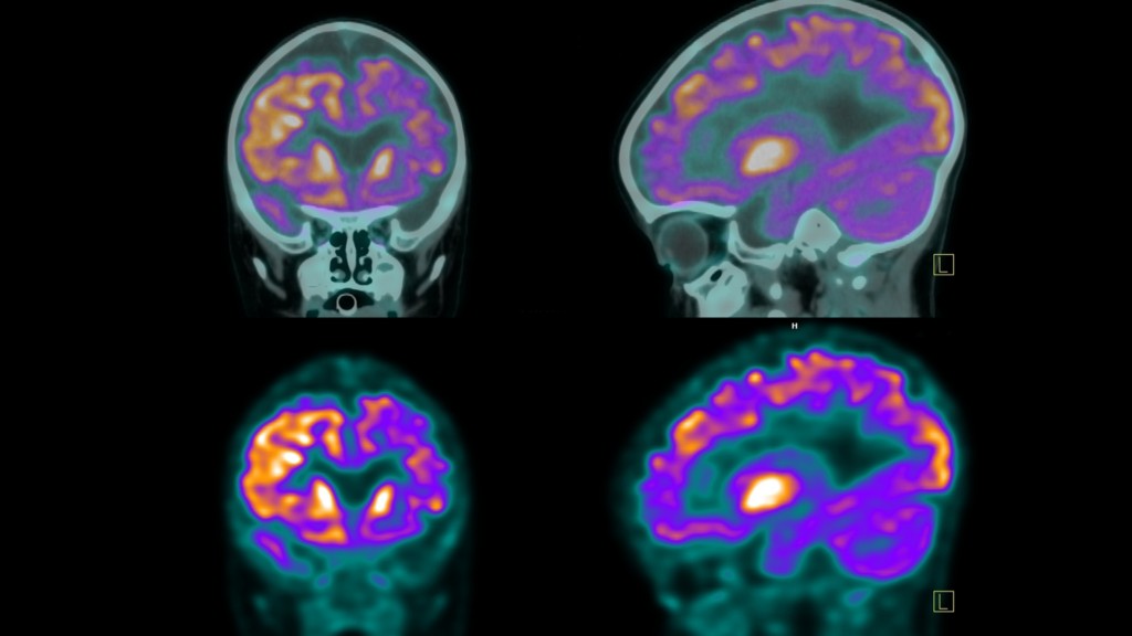 PET scan showing FDG uptake in the brain
2. Key Terminology in PET Scan Interpretation
Understanding the terminology used in PET scan reports is essential for interpreting the results effectively. Here are some key terms you may encounter:
2.1. Axial, Sagittal, and Coronal Planes
PET scanners capture images along three different planes to provide a comprehensive view of the body’s internal structures:
- Axial: Also known as the transverse view, this horizontal plane divides the body into top and bottom halves.
- Sagittal: The side view, which provides lateral images of the left and right halves of the body.
- Coronal: The face-on view, which creates images of the front and back halves of the body.
These planes help radiologists visualize and analyze the location and extent of any abnormalities.
2.2. Radiotracer
The radioactive substance used in PET scans is called a radiotracer. Fluorodeoxyglucose (FDG) is a common radiotracer, but others may be used depending on the specific clinical application. Your PET scan report will indicate which radiotracer was used, how much was administered, and the method of administration.
2.3. FDG Uptake
FDG uptake refers to the amount of radiotracer absorbed by cells. Different cell types have unique metabolic needs, so FDG will cluster at varying concentrations in different areas of the body. High FDG uptake generally indicates high metabolic activity, while low uptake suggests lower activity.
3. Decoding FDG Uptake: What It Means for Your Pet
FDG, or Fluorodeoxyglucose, is a glucose analog widely used as a radiotracer in PET scans. Understanding FDG uptake is crucial for interpreting the scan results and assessing your pet’s metabolic health.
3.1. What is FDG?
FDG is a radioactive tracer that mimics glucose, the primary source of energy for cells. Because cancer cells typically have higher metabolic rates than normal cells, they tend to absorb more FDG, making them visible on PET scans.
3.2. What Does FDG Uptake Indicate?
FDG uptake measures how much of the radiotracer is absorbed by cells. The level of uptake can provide valuable information about the metabolic activity of tissues and organs.
- High FDG Uptake: Indicates increased metabolic activity, which may be due to cancer, inflammation, infection, or other conditions.
- Low FDG Uptake: Suggests decreased metabolic activity, which may be normal for certain tissues or indicate tissue damage, necrosis, or successful treatment.
3.3. Factors Influencing FDG Uptake
Several factors can influence FDG uptake, including:
- Cell Type: Different cell types have varying metabolic needs.
- Disease State: Cancer, inflammation, and infection can increase metabolic activity.
- Treatment Effects: Successful treatments can reduce metabolic activity in diseased tissues.
- Technical Factors: The sensitivity of the PET scanner and the quality of the radiotracer can affect the results.
According to research from University of California, San Francisco, in 2024, monitoring FDG uptake helps to assess treatment response in cancer patients.
4. Interpreting Different Levels of FDG Uptake
Understanding the significance of different levels of FDG uptake is essential for accurate diagnosis and treatment planning.
4.1. No Uptake
No FDG uptake means the radiotracer isn’t being absorbed by the cells in the area being studied. This can have various implications, depending on the tissue type and clinical context:
- Tumor or Growth is Not Active: No uptake could indicate that a previously active tumor or growth is no longer metabolically active, possibly due to successful treatment.
- Normal Tissue: Certain tissues and areas of the body are expected to have low metabolic activity, making no uptake a normal finding.
- Detection Problems: Technical issues with the scanning equipment may prevent the detection of FDG uptake.
4.2. Normal FDG Uptake
Normal FDG uptake indicates that tissues are functioning within expected metabolic parameters. Organs like the brain, liver, and spleen typically have higher FDG uptake due to their high metabolic demands.
4.3. Mild or Low FDG Uptake
Low or mild FDG uptake can be normal for less active tissues or those high in fat. However, in areas expected to be more metabolically active, it may indicate the need for closer inspection:
- Less Active Tissue: Could suggest that previously active areas are now necrotic or that a tumor is less active.
- Possible Tumor Detection: Certain tumors may exhibit lower FDG uptake.
- Technical Limitations: Scanning equipment issues may affect the results.
4.4. Increased FDG Uptake
Increased FDG uptake means that cells in a specific area are absorbing more of the radiotracer than surrounding tissues. This higher uptake typically appears as brighter or more intense spots on the scan.
- Cancer: Cancer cells are usually more active and consume more glucose, resulting in higher FDG uptake.
- Inflammation or Infection: Areas with active inflammation or infection show increased uptake due to heightened immune activity.
- Tissue Healing: Healing tissues may exhibit increased FDG uptake following surgery or injury.
- Benign Conditions: Some non-cancerous growths can also show higher FDG uptake.
5. Understanding Standard Uptake Value (SUV)
SUV, or Standard Uptake Value, is a quantitative measure used in PET scans to determine the activity of the radiotracer in a specific area. It is a ratio that defines the concentration of the radiotracer at a specific point in time and is also known as the dose uptake ratio.
5.1. How is SUV Calculated?
The SUV is calculated by comparing the radiotracer concentration in a region of interest to the injected dose and the patient’s body weight. A higher SUV indicates increased metabolic activity.
5.2. SUV Interpretation
SUV values help in interpreting PET scan results, particularly when comparing multiple scans over time. Generally, metabolic activity is considered:
- Low Intensity: <5 SUV
- Moderate: 5-10 SUV
- Intense: 10-15 SUV
- Very Intense: >15 SUV
5.3. Clinical Significance of SUV
Changes in SUV values can provide valuable information about the progression of conditions or the effectiveness of treatments. An increasing SUV may indicate disease progression, while a decreasing SUV suggests a positive response to treatment.
6. Physiological Uptake vs. Abnormal Uptake
Distinguishing between physiological uptake and abnormal uptake is crucial for accurate PET scan interpretation.
6.1. What is Physiological Uptake?
Physiological uptake refers to the normal, expected absorption of a radiotracer in various organs and tissues. Different organs have different metabolic activities, leading to varying levels of radiotracer uptake.
- Liver: Normal metabolic activity results in uniform or mildly increased uptake.
- Kidneys and Bladder: Radiotracer is filtered by the kidneys and accumulates in the bladder, leading to normal uptake.
6.2. Differentiating Physiological Uptake from Abnormal Uptake
Radiologists use their expertise and knowledge of normal physiological patterns to differentiate normal uptake from abnormal uptake, which may indicate disease. Abnormal uptake patterns require further investigation to determine the underlying cause.
7. Metabolic Activity on a PET Scan: What It Reveals
Metabolic activity on a PET scan reflects how actively cells in the body are using glucose or other metabolic substrates. PET scans detect this activity using radiotracers, highlighting areas with increased metabolic uptake.
7.1. No Metabolic Activity
No metabolic activity in certain areas may be normal, but if unexpected, it can suggest:
- Damaged Tissue: Tissue may be necrotic or damaged.
- Blockages: Reduced blood flow or blockages may be present.
- Successful Treatment: In the context of a tumor, it may indicate a positive response to treatment.
7.2. Low-Grade or Mild Metabolic Activity
Low-grade metabolic activity may be normal for certain tissues, but if abnormal, it can indicate:
- Early Stages of Infections: Initial phases of tumors or infections.
- Scar Tissue: Fibrous tissues from surgeries or injuries.
- Aging: Natural decline in metabolic rate.
- Metabolic Disorders: Conditions like diabetes or hypothyroidism.
- Beginning of Successful Treatment/Healing: Reduction in metabolic activity as treatment takes effect.
7.3. Increased Metabolic Activity (Hypermetabolic)
Increased metabolic activity often indicates that cells are more active than normal, which can be a sign of inflammation, infection, or cancer. These areas show increased radiotracer uptake.
- Cancerous Tumors: Rapidly dividing cancer cells consume more glucose.
- Inflammation and Infection: Active immune responses increase cellular activity.
- Healing and Tissue Repair: Regenerating tissues post-injury show temporary increases.
- Benign Conditions: Some non-cancerous conditions may appear hypermetabolic.
According to a study by the National Institutes of Health in 2023, increased metabolic activity is a crucial marker for monitoring cancer progression and treatment response.
8. The Deauville Score: A Key Metric in Lymphoma Treatment
The Deauville score is an internationally recommended standard for reporting FDG uptake in treatment trials for Hodgkin’s and non-Hodgkin’s lymphomas. It is a visual interpretation that compares uptake in affected areas to uptake in the liver and mediastinum.
8.1. Understanding the Deauville Scale
The Deauville score runs from 1 to 5:
- No uptake
- Slight uptake, equal to or below uptake in the mediastinum
- Uptake above the mediastinum but below the liver
- Uptake slightly or moderately above the liver
- Noticeably increased uptake compared to the liver
8.2. Interpreting Deauville Scores
Lower numbers are better; 1 and 2 are considered complete responses, 3 is adequate, while 4 and 5 are considered inadequate.
9. Navigating Your Pet’s PET Scan Results: What to Expect
Understanding what to expect when reviewing your pet’s PET scan results can help you feel more prepared and empowered.
9.1. Unremarkable Findings
In medical terminology, “unremarkable” is a good thing; it means your PET scan reports no abnormal findings.
9.2. Accessing and Understanding Your Pet’s Report with PETS.EDU.VN
PETS.EDU.VN offers resources to help you understand your pet’s PET scan report. Here’s how we can assist:
- Comprehensive Information: Detailed explanations of medical terms and PET scan procedures.
- Expert Insights: Guidance on interpreting results and understanding their implications.
- Support and Resources: Access to additional information and support for pet owners.
10. Frequently Asked Questions (FAQs) About Uptake on PET Scans
10.1. What does “uptake” specifically refer to in a PET scan context?
In a PET scan, “uptake” refers to the absorption of a radioactive tracer, like FDG, by cells in the body. This uptake indicates the metabolic activity of those cells, providing valuable information about tissue and organ function.
10.2. How is FDG uptake measured during a PET scan?
FDG uptake is measured by the PET scanner, which detects the positrons emitted by the radioactive tracer. The scanner converts these emissions into detailed images that show the concentration of FDG in different areas of the body.
10.3. What is the significance of high FDG uptake in cancer diagnosis?
High FDG uptake is significant in cancer diagnosis because cancer cells typically have higher metabolic rates and consume more glucose than normal cells. This increased glucose consumption leads to higher FDG uptake, making cancer cells visible on PET scans.
10.4. Can non-cancerous conditions also cause increased FDG uptake?
Yes, non-cancerous conditions such as inflammation, infection, and tissue healing can also cause increased FDG uptake. These conditions increase cellular activity, leading to higher glucose consumption and FDG absorption.
10.5. How does SUV help in interpreting FDG uptake?
SUV, or Standard Uptake Value, is a quantitative measure that helps in interpreting FDG uptake by providing a standardized ratio of radiotracer concentration. This allows for a more objective assessment of metabolic activity and helps compare results across different scans.
10.6. What does it mean if a PET scan report mentions “physiological uptake”?
“Physiological uptake” refers to the normal, expected absorption of a radiotracer in various organs and tissues due to their natural metabolic activities. This is different from abnormal uptake, which may indicate disease.
10.7. How do doctors differentiate between normal and abnormal FDG uptake?
Doctors differentiate between normal and abnormal FDG uptake by considering factors such as the location, intensity, and pattern of uptake, as well as the patient’s medical history and other clinical findings.
10.8. What steps are taken if abnormal FDG uptake is detected?
If abnormal FDG uptake is detected, further testing is typically conducted to determine the underlying cause. This may include additional imaging studies, biopsies, or other diagnostic procedures.
10.9. How does the Deauville score relate to FDG uptake in lymphoma treatment?
The Deauville score is an internationally recognized standard for reporting FDG uptake in lymphoma treatment trials. It provides a visual interpretation of FDG uptake, helping to assess treatment response and determine the need for further intervention.
10.10. Where can I find reliable information about PET scans and FDG uptake for my pet?
You can find reliable information about PET scans and FDG uptake for your pet at PETS.EDU.VN. We offer comprehensive resources, expert insights, and support to help you understand your pet’s health and make informed decisions.
Conclusion: Empowering You with Knowledge for Informed Healthcare Decisions
Understanding the nuances of “uptake” on a PET scan is vital for making informed decisions about your pet’s health. At PETS.EDU.VN, we are committed to providing you with the knowledge and resources necessary to navigate complex medical information with confidence.
We understand that navigating the complexities of pet health can be overwhelming. That’s why PETS.EDU.VN is here to help. Our website offers comprehensive information, expert insights, and a supportive community to guide you every step of the way.
Ready to take control of your pet’s health journey? Visit pets.edu.vn today to explore our resources, connect with experts, and find the services your pet needs. For personalized assistance, contact us at 789 Paw Lane, Petville, CA 91234, United States, or reach out via WhatsApp at +1 555-987-6543. Your pet’s well-being is our priority.