Is a PET scan better than an MRI? Understanding the nuances between Positron Emission Tomography (PET) and Magnetic Resonance Imaging (MRI) is crucial for informed healthcare decisions. At PETS.EDU.VN, we empower you with detailed insights into these advanced imaging techniques, ensuring you’re well-prepared to discuss your options with your healthcare provider. This comprehensive guide explores the functionalities, advantages, and limitations of both PET and MRI scans, offering clarity on when one might be preferred over the other, enhancing diagnostic accuracy and patient care. Explore PETS.EDU.VN for more resources on diagnostic imaging and pet health solutions.
1. Understanding PET and MRI Scans
Different types of imaging tests play a vital role in diagnosing various medical conditions, including X-rays, ultrasound, CT scans, MRI, and PET scans. Each of these imaging modalities offers unique advantages and provides specific information about the body. Understanding the differences between them is crucial for healthcare professionals and patients alike.
1.1. Key Differences Between PET and MRI Scans
Here’s a comparison highlighting the core distinctions between PET and MRI scans:
| Feature | PET Scan | MRI Scan |
|---|---|---|
| Function | Assesses cellular-level energy use in real-time to detect abnormalities. | Uses strong magnets and radio waves to create detailed, static images of body structures. |
| Method | Employs a radioactive tracer, typically fluorodeoxyglucose (FDG). | Can be performed with or without contrast dye to enhance image clarity. |
| Radiation | Involves exposure to a small amount of radiation from the tracer. | Does not use radiation, making it a safer option in certain scenarios. |
| Tissue Focus | Detects tissue changes at the cellular level, sometimes before MRI can visualize them. | Excels at visualizing soft tissues like muscles, fat, and organs. |
| Image Type | Provides functional images showing metabolic activity. | Delivers high-resolution anatomical images. |
| Scan Duration | Typically takes 30 minutes to 1 hour. | Can range from 15 to 90 minutes, depending on the body area and complexity of the scan. |
| Contrast Agent | Radioactive tracer (e.g., FDG) | Gadolinium-based contrast agents (if needed) |
| Primary Use | Detecting cancers, neurological disorders, and cardiovascular diseases. | Imaging brain, spine, joints, soft tissues, and internal organs. |
| Claustrophobia | Generally less claustrophobic due to shorter scan times and more open design. | Can be challenging for those with claustrophobia, although open MRI options are available. |
| Metal Implants | Less affected by metal implants compared to MRI. | Requires careful screening for metal implants due to strong magnetic field. |

1.2. How PET Scans Work
A PET scan is a sophisticated nuclear medicine imaging technique that generates 3D images, revealing how well different parts of the body function. About an hour before the scan, a small amount of radioactive tracer is injected into the patient’s vein. This tracer emits positrons, which the scanner detects to identify areas of the body using the most energy.
1.3. The Tracer and Its Role
The most common tracer is fluorodeoxyglucose (FDG), which is similar to glucose (sugar). Cancerous areas and other abnormalities consume glucose at a much higher rate than normal cells, making them easily detectable. Doctors consider factors like diabetes and blood sugar levels when interpreting PET scan results. The advantage of PET scans is their ability to detect changes at the cellular level, often before MRI or CT scans can identify them. However, follow-up MRI or CT scans may be needed to confirm whether the changes are cancerous.
1.4. The PET Scan Procedure
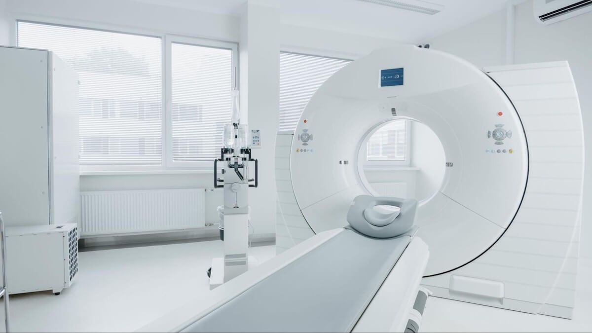
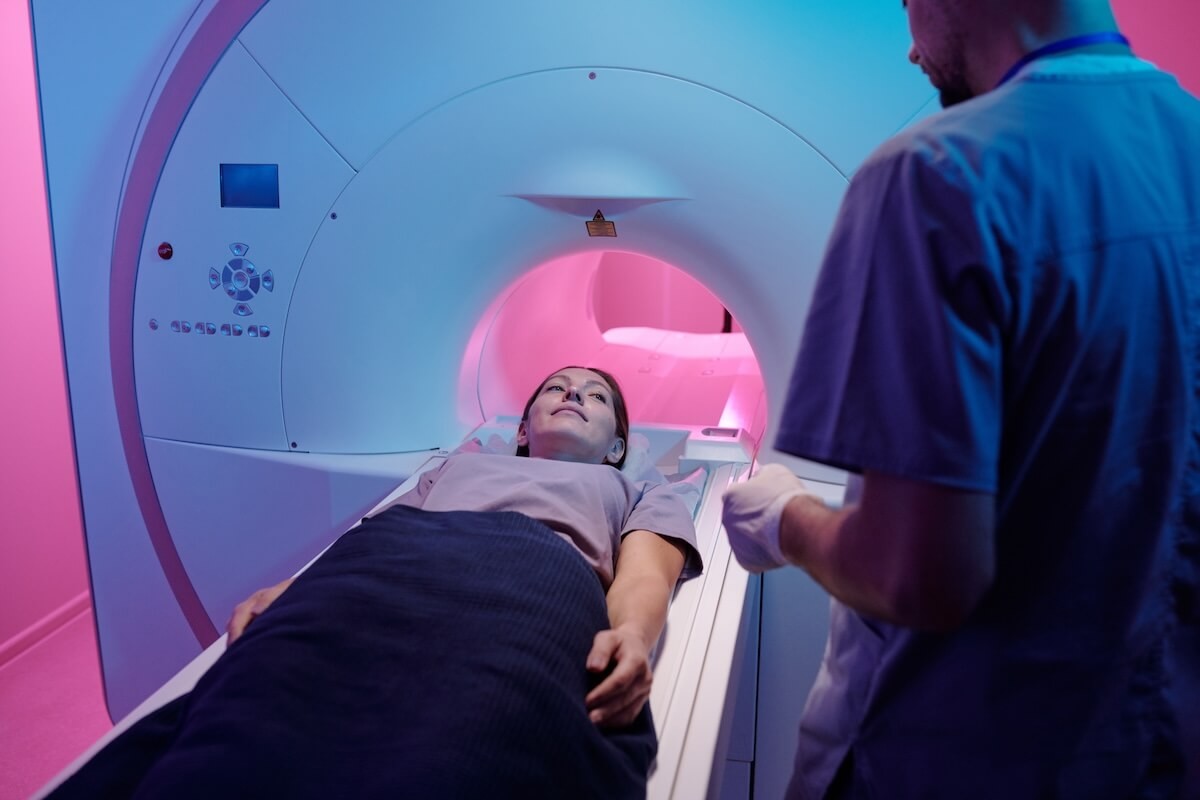
During the scan, the patient lies on their back on a flat bed that slides into a donut-shaped PET scanner. The scan is painless and usually lasts between 30 minutes to one hour. The PET scanner captures the distribution of the radioactive tracer, creating detailed images of the body’s metabolic activity. This information helps healthcare professionals diagnose and monitor various conditions, including cancer, heart disease, and neurological disorders.
1.5. How MRI Scans Work
MRI is a non-invasive imaging technique that uses radio waves and strong magnets to create detailed images of the inside of the body. The technology harnesses a powerful computer to transform these radio waves and magnetic fields into highly detailed images.
1.6. MRI with and without Contrast
MRI scans can take between 15 to 90 minutes, depending on the complexity of the scan and the area being examined. They can be performed with or without contrast agents. While most soft tissues can be visualized without contrast, contrast agents like gadolinium can enhance image clarity in certain scenarios.
1.7. The MRI Procedure
In a contrast MRI, the contrast dye is typically injected into the patient’s vein before the scan. The patient lies on their back on a flat bed, which is then moved into the MRI scanner. The MRI machine uses strong magnetic fields and radio waves to generate cross-sectional images of the body. These images provide detailed views of soft tissues, organs, and other structures, aiding in the diagnosis and monitoring of various medical conditions.
Once a PET or MRI scan is completed, a radiologist or nuclear medicine physician reviews the scans and sends their findings to the referring healthcare practitioner. The practitioner then shares the results with the patient during a follow-up appointment, explaining the findings and discussing any necessary treatment plans.
2. What Can PET and MRI Scans Show?
PET and MRI scans are invaluable diagnostic tools, each capable of revealing different aspects of the body’s condition. The choice between a PET scan and an MRI depends on the specific clinical needs and the suspected conditions being investigated. Let’s delve into the capabilities of each.
2.1. PET Scan Applications
A PET scan can be a full-body scan or focus on a specific area, aiding in the diagnosis of various conditions:
- Cancer Detection and Staging: PET scans are highly effective in detecting different types of cancer, determining if cancer has spread (metastasis), and evaluating the effectiveness of cancer treatments. The scan identifies areas with high metabolic activity, which can indicate cancerous cells.
- Brain Disorders: PET scans can help diagnose brain disorders such as Alzheimer’s disease, brain tumors, and epilepsy. By measuring glucose metabolism in the brain, PET scans can detect abnormalities associated with these conditions.
- Heart Conditions: PET scans can identify heart conditions like coronary artery disease by detecting areas of decreased blood flow. This helps in assessing the extent of heart damage and planning appropriate interventions.
2.1.1. Advantages of PET Scans in Cancer Diagnosis
PET scans are particularly useful in oncology for several reasons:
- Early Detection: PET scans can detect metabolic changes in cells before structural changes are visible on other imaging modalities like CT or MRI.
- Whole-Body Assessment: PET scans can scan the entire body in a single session, allowing for comprehensive assessment of cancer spread.
- Treatment Monitoring: PET scans can monitor the response of cancer to treatment, helping healthcare professionals adjust treatment plans as needed.
2.1.2. Limitations of PET Scans in Cancer Diagnosis
Despite their advantages, PET scans have some limitations:
- Not All Cancers Show Up: Some cancers may not be visible on PET scans due to their low metabolic activity.
- False Positives: Non-cancerous conditions, such as infections or inflammation, can also cause increased metabolic activity, leading to false positive results.
- Need for Additional Imaging: In some cases, additional imaging or tests may be needed to confirm if an area picked up by the tracer is cancerous.
2.2. MRI Scan Applications
MRI is versatile and particularly adept at imaging soft tissues. It can be used as a full-body scan or focused on a specific area to investigate a range of conditions:
- Brain and Spine: MRI is used to diagnose conditions such as multiple sclerosis, stroke, aneurysms, and tumors of the brain and spinal cord. Its high-resolution imaging provides detailed views of these structures.
- Bones and Joints: MRI can identify primary bone tumors, joint injuries, and tendon injuries. It provides detailed images of the musculoskeletal system, aiding in accurate diagnosis and treatment planning.
- Soft Tissues: MRI excels at visualizing organs, muscles, nerves, tendons, and ligaments, including soft tissue sarcomas. This makes it invaluable for diagnosing soft tissue injuries and abnormalities.
- Abdominal and Pelvic Organs: MRI can visualize the liver, kidneys, and pancreas, and detect tumors in the prostate, bladder, uterus, or ovaries. This is crucial for diagnosing and staging cancers and other conditions affecting these organs.
- Heart and Lungs: MRI can identify structural and functional abnormalities, tumors, and diseases affecting the heart muscle. Cardiac MRI is used to assess heart function, detect scar tissue, and diagnose congenital heart defects.
2.2.1. Specialized MRI Techniques
- Breast MRI: A dedicated breast MRI with a specialized breast coil can be performed to detect breast cancer and assess the extent of disease. It is often used in women with a high risk of breast cancer or those with dense breast tissue.
- Magnetic Resonance Angiography (MRA): MRA measures blood flow through blood vessels, aiding in the diagnosis of vascular conditions such as aneurysms and arterial blockages.
2.2.2. Whole-Body MRI Screening
Whole-body MRI scans are available for asymptomatic individuals who want to proactively monitor their health. These scans can detect early signs of disease, allowing for timely intervention and treatment.
PET and MRI scans offer distinct but complementary capabilities, providing comprehensive diagnostic information across a wide range of medical conditions. While PET excels at detecting metabolic activity associated with cancer, brain disorders, and heart conditions, MRI provides detailed anatomical images of soft tissues, organs, and the musculoskeletal system.
3. PET Scan vs. MRI: Which Is More Accurate?
When it comes to diagnostic imaging, accuracy is paramount. However, determining whether a PET scan or an MRI is more accurate depends largely on the specific condition being investigated. PETS.EDU.VN emphasizes that these technologies are not mutually exclusive but rather complementary, often used in conjunction to provide a comprehensive understanding of a patient’s health.
3.1. Complementary Use of PET and MRI
In many clinical scenarios, the most effective approach involves using both PET and MRI scans. For example, a PET-MRI scan combines the functional insights of PET with the detailed anatomical imaging of MRI. This combination is particularly useful in oncology, where it can help confirm whether an abnormality seen on a PET scan is cancerous (malignant).
3.2. Situations Favoring PET Scans
PET scans excel in detecting metabolic activity, making them highly accurate for:
- Cancer Detection and Staging: PET scans can identify cancerous cells by detecting their increased glucose metabolism, even before structural changes are visible on MRI.
- Neurological Disorders: PET scans can measure brain metabolism, helping diagnose conditions like Alzheimer’s disease and epilepsy.
- Cardiovascular Disease: PET scans can assess blood flow and myocardial viability, aiding in the diagnosis of coronary artery disease.
3.3. Situations Favoring MRI Scans
MRI scans provide high-resolution anatomical images, making them highly accurate for:
- Brain and Spinal Cord Imaging: MRI is the gold standard for imaging the brain and spinal cord, detecting conditions like tumors, multiple sclerosis, and stroke.
- Musculoskeletal Imaging: MRI is excellent for visualizing soft tissues, including muscles, ligaments, tendons, and cartilage, making it ideal for diagnosing joint and soft tissue injuries.
- Abdominal and Pelvic Imaging: MRI can provide detailed images of the liver, kidneys, pancreas, and other abdominal and pelvic organs, helping diagnose tumors, cysts, and other abnormalities.
3.4. Case Studies and Examples
To illustrate the complementary nature of PET and MRI, consider the following examples:
- Lung Cancer: A PET scan may detect a suspicious nodule in the lung, while an MRI can provide detailed information about its size, shape, and location, helping determine the stage of cancer and guide treatment decisions.
- Brain Tumor: An MRI can identify a brain tumor, while a PET scan can assess its metabolic activity, helping differentiate between high-grade and low-grade tumors and guide surgical planning.
- Cardiac Disease: An MRI can assess the structure and function of the heart, while a PET scan can measure blood flow to the heart muscle, helping diagnose coronary artery disease and assess myocardial viability.
4. Potential Risks and Challenges
Both PET and MRI scans are considered safe and non-invasive (excluding the injection of the tracer or MRI contrast). However, it’s crucial to be aware of potential risks, side effects, and challenges associated with each.
4.1. Radiation Exposure from PET Scans
One of the primary concerns with PET scans is exposure to radiation. Although the amount of radiation is small, it’s essential to understand the associated risks. According to the National Cancer Institute, the risk of developing cancer from the low-dose radiation used in PET scans is very small.
4.1.1. Minimizing Radiation Exposure
- Hydration: Drinking plenty of water after the scan helps flush the radioactive tracer out of the body more quickly.
- Limited Contact: Patients should avoid close contact with pregnant women, young children, and babies for about six hours after the scan.
4.1.2. Risks of Repeated PET Scans
The small risk from radiation exposure increases with repeated PET scans, especially when combined with CT scans, which also involve radiation. However, the overall risks remain very small.
4.2. MRI and the Absence of Radiation
MRI scans do not involve radiation exposure, making them a preferred option for patients who require frequent imaging or those who are particularly sensitive to radiation, such as pregnant women.
4.3. Pregnancy Considerations
The American College of Obstetricians and Gynecologists recommends that MRI scans during pregnancy should be considered on a case-by-case basis, after careful discussion with a healthcare practitioner. MRI scans without contrast are generally preferred. PET scans are typically avoided during pregnancy and breastfeeding due to the risk of radiation exposure to the fetus or infant.
4.4. Claustrophobia
Both PET and MRI scans require patients to lie still inside a scanner, which can be challenging for those with claustrophobia.
4.4.1. Managing Claustrophobia
- Sedation: A mild sedative can be requested before the scan to help patients relax.
- Open MRI: Open MRI machines are available, offering a more spacious environment. However, the images may be lower resolution due to a weaker magnetic field.
PET scans are generally easier to manage for claustrophobic patients because the scanner is shorter, and the bed moves in and out during the scan.
4.5. The Need to Lie Still
Remaining still for the duration of the scan is crucial for obtaining clear images. This can be uncomfortable, especially for longer MRI scans. Patients should discuss any concerns with their healthcare provider before the scan.
4.6. Metal Implants and MRI
The strong magnetic field used in MRI scans requires the removal of all metal items. MRI scans cannot be performed on patients with certain types of metal inside their bodies, including:
- Cochlear implants
- Pacemakers or implantable defibrillators
- Metal fragments from injuries or dental bridges
Patients are given a detailed checklist and consent form to complete before the scan to determine if it is safe to proceed.
4.7. Allergic Reactions
Allergic reactions to the contrast dye used in MRI or the radiotracer used in PET scans are rare. Patients with known allergies should inform their healthcare practitioner before the scan. Side effects from MRI contrast dye can include headache, dizziness, nausea, vomiting, or skin rash. Rarely, swelling and pain may occur around the injection site of the PET radiotracer.
4.8. Noise Levels
Both PET and MRI scans can be noisy. Earplugs or headphones are typically provided to protect hearing and provide a distraction. Technicians can communicate with patients during the scan to alleviate any anxiety.
5. Making the Right Choice for Your Situation
Choosing between a PET scan and an MRI scan depends on the specific diagnostic needs and clinical context. PETS.EDU.VN is committed to providing you with the information you need to make informed decisions, in consultation with your healthcare provider.
5.1. Diagnostic Purposes
When imaging is needed for diagnostic purposes, the healthcare practitioner, in consultation with the patient, will make a decision based on clinical needs and discuss the risks and benefits of each option. If there’s an urgent need for a scan, the benefits of ruling out a serious condition typically outweigh any potential risks.
5.2. Proactive Health Screening
For individuals considering proactive health screening, MRI is an excellent choice due to its non-invasive nature and lack of radiation exposure.
5.3. Factors Influencing the Choice
Several factors influence the decision between PET and MRI scans:
- Clinical Indication: The specific condition being investigated is the primary determinant. PET scans are preferred for detecting metabolic activity, while MRI is superior for anatomical imaging.
- Patient Factors: Patient-specific factors such as pregnancy, claustrophobia, presence of metal implants, and allergies to contrast agents must be considered.
- Availability and Cost: The availability and cost of each imaging modality can also influence the decision.
5.4. Summary Table: PET Scan vs. MRI
| Feature | PET Scan | MRI Scan |
|---|---|---|
| Primary Use | Detecting cancers, neurological disorders, and cardiovascular diseases by assessing metabolic activity. | Imaging brain, spine, joints, soft tissues, and internal organs with high-resolution anatomical detail. |
| Radiation | Involves exposure to a small amount of radiation. | Does not use radiation. |
| Contrast Agent | Radioactive tracer (e.g., FDG). | Gadolinium-based contrast agents (if needed). |
| Scan Duration | Typically 30 minutes to 1 hour. | Can range from 15 to 90 minutes. |
| Claustrophobia | Generally less claustrophobic. | Can be challenging for those with claustrophobia; open MRI options are available. |
| Metal Implants | Less affected by metal implants. | Requires careful screening for metal implants. |
| Accuracy | Highly accurate for detecting metabolic changes at the cellular level. | Highly accurate for visualizing anatomical structures and soft tissues. |
| Best Suited For | Early detection of cancer, monitoring treatment response, diagnosing brain disorders, and heart conditions. | Diagnosing brain and spinal cord conditions, musculoskeletal injuries, abdominal and pelvic disorders, and heart conditions. |
Understanding the unique strengths and limitations of PET and MRI scans empowers patients and healthcare providers to make informed decisions about diagnostic imaging. PETS.EDU.VN is dedicated to providing comprehensive information and resources to help you navigate the complexities of pet health.
6. Innovations and Future Trends in PET and MRI Technology
The fields of PET and MRI technology are continually evolving, with ongoing research and development leading to significant advancements. These innovations promise to improve diagnostic accuracy, reduce scan times, and enhance patient comfort. Here, we explore some of the cutting-edge developments and future trends in PET and MRI technology.
6.1. Advancements in PET Technology
Recent advancements in PET technology are focused on improving image resolution, reducing radiation exposure, and expanding the range of detectable tracers.
- Improved Image Resolution: New PET scanners are equipped with advanced detectors and reconstruction algorithms that provide higher resolution images. This allows for the detection of smaller lesions and more accurate staging of cancer.
- Reduced Radiation Exposure: Efforts are being made to reduce the amount of radioactive tracer needed for PET scans. This is achieved through the development of more sensitive detectors and optimized scanning protocols.
- Novel Tracers: Researchers are developing new tracers that target specific biological processes, such as inflammation, angiogenesis, and cell proliferation. These tracers can provide valuable insights into the pathophysiology of various diseases.
- PET/MRI Hybrid Imaging: The integration of PET and MRI technologies into a single hybrid scanner allows for simultaneous acquisition of functional and anatomical images. This provides a more comprehensive assessment of disease and can improve diagnostic accuracy.
6.2. Advancements in MRI Technology
MRI technology is also advancing rapidly, with a focus on improving image quality, reducing scan times, and enhancing patient comfort.
- Higher Field Strengths: MRI scanners with higher magnetic field strengths (e.g., 3 Tesla and 7 Tesla) provide improved signal-to-noise ratio and higher resolution images. This allows for the detection of subtle abnormalities and more detailed visualization of anatomical structures.
- Compressed Sensing: Compressed sensing is a technique that allows for faster MRI scans by acquiring fewer data points. This can significantly reduce scan times and improve patient comfort, particularly for those who have difficulty lying still.
- Artificial Intelligence (AI): AI algorithms are being developed to enhance MRI image quality, automate image analysis, and improve diagnostic accuracy. AI can also be used to personalize MRI protocols based on individual patient characteristics.
- Open MRI: Open MRI scanners offer a more spacious and comfortable environment for patients, particularly those with claustrophobia. While open MRI scanners typically have lower magnetic field strengths than closed MRI scanners, advancements in technology are improving their image quality.
6.3. Future Trends in PET and MRI Technology
Several exciting trends are shaping the future of PET and MRI technology.
- Molecular Imaging: Molecular imaging, which uses PET and MRI to visualize biological processes at the molecular level, is becoming increasingly important in personalized medicine. This allows for the development of targeted therapies that are tailored to individual patient characteristics.
- Multi-Modal Imaging: Combining PET and MRI with other imaging modalities, such as CT and ultrasound, can provide a more comprehensive assessment of disease. This multi-modal approach is particularly useful in oncology, where it can help guide treatment decisions.
- Quantitative Imaging: Quantitative imaging techniques, which measure the absolute values of PET and MRI parameters, are becoming increasingly important in clinical research and drug development. This allows for more accurate assessment of treatment response and disease progression.
- Point-of-Care Imaging: The development of portable and point-of-care PET and MRI scanners could revolutionize medical imaging, making it more accessible and affordable, especially in underserved communities.
6.4. Benefits of Technological Advancements
The advancements in PET and MRI technology offer several benefits:
- Improved Diagnostic Accuracy: Higher resolution images and novel tracers allow for the detection of smaller lesions and more accurate staging of disease.
- Reduced Scan Times: Faster scan times improve patient comfort and increase throughput.
- Enhanced Patient Comfort: Open MRI scanners and other comfort-enhancing features make imaging more accessible to patients with claustrophobia and other concerns.
- Personalized Medicine: Molecular imaging and quantitative imaging techniques enable personalized treatment strategies based on individual patient characteristics.
7. Understanding the Costs Associated with PET and MRI Scans
Navigating the financial aspects of medical imaging can be a significant concern for many patients. Understanding the costs associated with PET and MRI scans, as well as the factors that influence these costs, is essential for making informed decisions about your healthcare. PETS.EDU.VN aims to provide clarity on this topic, helping you to better plan and manage your medical expenses.
7.1. Factors Influencing the Cost of PET and MRI Scans
Several factors can affect the cost of PET and MRI scans:
- Geographic Location: The cost of medical imaging can vary significantly depending on the region and even within different facilities in the same city. Areas with higher costs of living typically have higher medical expenses.
- Type of Facility: Hospitals, outpatient imaging centers, and private clinics may have different pricing structures. Generally, scans performed in hospitals tend to be more expensive than those in outpatient centers due to higher overhead costs.
- Type of Scan: The specific type of PET or MRI scan required can impact the cost. For instance, a full-body PET scan may cost more than a localized scan. Similarly, MRI scans with contrast agents or specialized sequences may also increase the price.
- Insurance Coverage: The extent of your insurance coverage plays a crucial role in determining your out-of-pocket expenses. It is essential to check with your insurance provider to understand what portion of the scan will be covered and if any pre-authorization is required.
- Physician Fees: The cost of the radiologist’s interpretation of the scan is often separate from the facility fee. These professional fees can vary based on the radiologist’s experience and specialization.
7.2. Average Costs of PET and MRI Scans
While the exact cost can vary widely, here are some general estimates for the average costs of PET and MRI scans in the United States:
- PET Scan: The average cost of a PET scan ranges from $2,000 to $5,000 per scan.
- MRI Scan: The average cost of an MRI scan ranges from $400 to $3,500 per scan.
It’s important to note that these are just estimates, and the actual cost may be higher or lower depending on the factors mentioned above.
7.3. Insurance Coverage for PET and MRI Scans
Most health insurance plans, including Medicare and Medicaid, cover PET and MRI scans when they are deemed medically necessary by a healthcare provider. However, coverage can vary depending on your specific plan and the reason for the scan.
- Pre-Authorization: Many insurance plans require pre-authorization before a PET or MRI scan can be performed. This involves your healthcare provider submitting documentation to the insurance company to justify the medical necessity of the scan.
- Deductibles and Co-pays: Even if your insurance covers the scan, you may still be responsible for meeting your deductible or paying a co-pay. The deductible is the amount you must pay out-of-pocket before your insurance begins to cover medical expenses, while a co-pay is a fixed amount you pay for each service.
- Out-of-Network Costs: If you choose to have a PET or MRI scan performed at an out-of-network facility, your insurance may cover a smaller portion of the cost, leaving you with higher out-of-pocket expenses.
7.4. Strategies to Manage the Costs of PET and MRI Scans
Here are some strategies to help manage the costs of PET and MRI scans:
- Check Insurance Coverage: Contact your insurance provider to understand your coverage for PET and MRI scans, including any pre-authorization requirements, deductibles, and co-pays.
- Compare Prices: Call different imaging facilities to compare prices for the specific scan you need. Ask about any discounts or payment plans that may be available.
- Choose In-Network Providers: Opt for imaging facilities that are within your insurance network to minimize out-of-pocket expenses.
- Ask About Bundled Pricing: Some facilities offer bundled pricing for the scan and radiologist interpretation, which may be more cost-effective than paying for each separately.
- Negotiate Payment Plans: If you are unable to afford the full cost of the scan upfront, ask the facility about payment plan options.
7.5. Financial Assistance Programs
Several financial assistance programs may help offset the costs of medical imaging:
- Hospital Financial Aid: Many hospitals offer financial aid programs to help patients with limited incomes afford medical care.
- Non-Profit Organizations: Some non-profit organizations provide financial assistance to patients who need help paying for medical expenses.
- Government Programs: Government programs such as Medicaid may cover the costs of PET and MRI scans for eligible individuals.
8. Ethical Considerations in the Use of PET and MRI Scans
The use of PET and MRI scans in medical diagnostics and research raises several ethical considerations that healthcare professionals, researchers, and patients must address. PETS.EDU.VN recognizes the importance of these ethical considerations and aims to promote responsible and ethical practices in the use of medical imaging technologies.
8.1. Justification of Scan Requests
One of the primary ethical considerations in the use of PET and MRI scans is the justification of scan requests. Healthcare providers must carefully evaluate the potential benefits and risks of each scan and ensure that the scan is medically necessary and appropriate for the patient’s condition.
- Clinical Necessity: PET and MRI scans should be ordered only when they are likely to provide valuable information that cannot be obtained through other, less invasive or less costly means.
- Appropriate Use Criteria: Healthcare providers should adhere to established guidelines and appropriate use criteria for PET and MRI scans to ensure that the scans are used judiciously and effectively.
- Patient Education: Patients should be informed about the reasons for the scan, the potential benefits and risks, and alternative diagnostic options.
8.2. Informed Consent
Obtaining informed consent from patients before performing a PET or MRI scan is essential for respecting patient autonomy and ensuring that patients have the information they need to make informed decisions about their healthcare.
- Disclosure of Risks and Benefits: Patients should be informed about the potential risks and benefits of the scan, including the risks of radiation exposure (in the case of PET scans), allergic reactions to contrast agents, and claustrophobia.
- Alternative Options: Patients should be informed about alternative diagnostic options and their associated risks and benefits.
- Voluntary Decision: Patients must have the freedom to choose whether or not to undergo the scan, without coercion or undue influence from healthcare providers.
8.3. Privacy and Confidentiality
Protecting the privacy and confidentiality of patient information is a fundamental ethical obligation for healthcare providers.
- Data Security: Healthcare providers must implement appropriate measures to protect patient data from unauthorized access, use, or disclosure.
- Data Sharing: Patient data should be shared only with individuals who have a legitimate need to know, and patients should be informed about how their data will be used and with whom it will be shared.
- Compliance with Regulations: Healthcare providers must comply with all applicable privacy laws and regulations, such as the Health Insurance Portability and Accountability Act (HIPAA).
8.4. Equity and Access
Ensuring equitable access to PET and MRI scans is an important ethical consideration, particularly in underserved communities.
- Affordability: Efforts should be made to make PET and MRI scans affordable for all patients, regardless of their socioeconomic status.
- Availability: PET and MRI scanners should be available in all geographic areas, including rural and underserved communities.
- Cultural Sensitivity: Healthcare providers should be sensitive to the cultural beliefs and values of their patients and provide culturally appropriate care.
8.5. Research Ethics
The use of PET and MRI scans in research raises additional ethical considerations.
- Scientific Validity: Research studies involving PET and MRI scans should be scientifically valid and designed to answer important research questions.
- Ethical Review: Research studies involving human subjects must be reviewed and approved by an institutional review board (IRB) to ensure that they are ethically sound and protect the rights and welfare of research participants.
- Informed Consent: Research participants must provide informed consent before participating in a research study, and they should be informed about the purpose of the study, the potential risks and benefits, and their right to withdraw from the study at any time.
9. Real-World Applications: Case Studies
To further illustrate the practical applications of PET and MRI scans, let’s explore some real-world case studies where these imaging techniques have played a crucial role in diagnosis, treatment planning, and monitoring.
9.1. Case Study 1: Lung Cancer Diagnosis and Staging
Patient: A 65-year-old male with a history of smoking presented with a persistent cough and shortness of breath.
Diagnostic Process:
- Initial Assessment: A chest X-ray revealed a suspicious nodule in the lung.
- PET Scan: A PET scan was performed to assess the metabolic activity of the nodule and determine if it was cancerous. The PET scan showed increased glucose uptake in the nodule, indicating a high likelihood of malignancy.
- MRI Scan: An MRI of the chest was performed to provide detailed anatomical information about the nodule, including its size, shape, and location. The MRI also helped to assess if the cancer had spread to nearby structures, such as the chest wall or mediastinum.
- Diagnosis: Based on the PET and MRI findings, the patient was diagnosed with Stage IIIA non-small cell lung cancer.
Treatment Planning: The PET and MRI scans were used to guide treatment planning. The patient underwent chemotherapy and radiation therapy, followed by surgical resection of the lung nodule.
Outcome: The patient responded well to treatment and has been in remission for five years.
9.2. Case Study 2: Brain Tumor Evaluation
Patient: A 40-year-old female presented with headaches, vision changes, and seizures.
Diagnostic Process:
- Initial Assessment: A neurological examination revealed several abnormalities, including papilledema (swelling of the optic disc) and visual field deficits.
- MRI Scan: An MRI of the brain was performed to evaluate the possibility of a brain tumor. The MRI revealed a mass in the left temporal lobe, consistent with a glioma (a type of brain tumor).
- PET Scan: A PET scan was performed to assess the metabolic activity of the tumor and determine its grade (aggressiveness). The PET scan showed increased glucose uptake in the tumor, indicating a high-grade glioma.
Treatment Planning: The PET and MRI scans were used to guide treatment planning. The patient underwent surgical resection of the tumor, followed by radiation therapy and chemotherapy.
Outcome: The patient’s symptoms improved after surgery, and she has been able to maintain a good quality of life with ongoing treatment.
9.3. Case Study 3: Cardiac Viability Assessment
Patient: A 70-year-old male with a history of coronary artery disease presented with chest pain and shortness of breath.
Diagnostic Process:
- Initial Assessment: An electrocardiogram (ECG) showed evidence of ischemia (reduced blood flow) in the heart.
- MRI Scan: An MRI of the heart was performed to assess the structure and function of the heart muscle. The MRI revealed areas of scar tissue in the left ventricle, indicating a prior heart attack.
- PET Scan: A PET scan was performed to assess the viability (health) of the heart muscle. The PET scan showed areas of reduced blood flow but preserved metabolism, indicating that the heart muscle was still viable and could potentially benefit from revascularization (restoring blood flow).
Treatment Planning: Based on the PET and MRI findings, the patient underwent coronary artery bypass grafting (CABG) to restore blood flow to the heart muscle.
Outcome: The patient’s symptoms improved after surgery, and his heart function improved.
10. PETS.EDU.VN: Your Partner in Pet Health
At pets.edu.vn, we understand the unique bond you share with your pets and the importance of providing them with the best possible care. While this article has focused on PET and MRI scans in human medicine, the principles and technologies are increasingly being applied in veterinary medicine to diagnose and treat various conditions in animals.
10.1. Cutting-Edge Veterinary Diagnostics
We are committed to providing cutting-edge veterinary diagnostics, including advanced imaging techniques like PET and MRI, to help our clients make informed decisions about their pets’ health.
- Expert Veterinary Specialists: Our team of expert veterinary specialists is trained in the interpretation of PET and MRI scans and can provide valuable insights into your pet’s condition.
- State-of-the-Art Imaging Facilities: We partner with state-of-the-art imaging facilities to provide your pet with the highest quality imaging services.