Are you curious about medical imaging and wondering, “Is Mri Better Than Pet Scan?” This comprehensive guide from PETS.EDU.VN explores the nuances of MRI and PET scans, offering a clear understanding of their differences, benefits, and ideal applications. Let’s delve into the world of medical imaging and discover which scan might be the right choice for specific diagnostic needs, helping you make informed decisions about your health or the health of your beloved pet. With PETS.EDU.VN, navigate the complexities of PET scans and magnetic resonance imaging, and uncover insights into diagnostic radiology and medical imaging technology.
1. Understanding the Core Differences Between PET and MRI Scans
When it comes to medical imaging, both PET (Positron Emission Tomography) and MRI (Magnetic Resonance Imaging) scans are powerful tools, but they operate on fundamentally different principles. Understanding these differences is key to appreciating when one might be preferred over the other. Let’s explore the distinct features that set these two imaging techniques apart.
-
PET Scan: Functional Imaging at the Cellular Level: A PET scan excels at showing how your body’s tissues and organs are functioning at a cellular level. It uses a radioactive tracer to detect metabolic activity, highlighting areas where cells are more active than usual, such as in tumors or areas of inflammation.
-
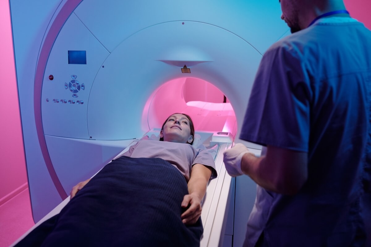
MRI Scan: Detailed Anatomical Imaging: In contrast, an MRI scan provides detailed, still images of the body’s anatomy using strong magnets and radio waves. It’s particularly adept at visualizing soft tissues like the brain, spinal cord, and joints.
alt: A patient undergoing an MRI scan, showcasing the detailed anatomical imaging capabilities.
1.1. Key Distinctions in Technology and Application
The fundamental technology behind each scan dictates its best use cases.
- PET Scan: Radioactive Tracers and Metabolic Activity: PET scans rely on the detection of radioactive tracers injected into the body. These tracers accumulate in areas of high metabolic activity, allowing doctors to see how well certain tissues or organs are functioning. This is particularly useful in detecting cancer, heart problems, and brain disorders.
- MRI Scan: Magnetic Fields and Radio Waves: MRI scans, on the other hand, use powerful magnetic fields and radio waves to generate detailed images of the body’s internal structures. They are excellent for visualizing soft tissues, making them invaluable in diagnosing conditions affecting the brain, spinal cord, joints, and other soft tissue areas.
- Radiation Exposure: A significant difference between the two is radiation exposure. PET scans involve a small amount of radiation due to the radioactive tracer, while MRI scans do not use radiation at all, making them a safer option for pregnant women and children when appropriate.
- Use of Contrast: Both scans can be enhanced with contrast agents. In PET scans, the tracer itself acts as the contrast. In MRI, substances like gadolinium can be injected to improve image clarity, especially when looking at blood vessels or inflammation.
- Timing: The timing of these scans also differs. A PET scan might reveal changes at a cellular level before they are visible on an MRI. However, MRIs can provide more detailed anatomical information to confirm the nature and extent of these changes.
Understanding these key differences helps healthcare professionals determine which scan is most appropriate for diagnosing specific conditions. Each scan provides unique information that can be crucial in making accurate diagnoses and guiding treatment decisions.
2. How PET and MRI Scans Work: A Detailed Look
To truly understand whether an MRI is better than a PET scan, it’s essential to delve into the mechanics of how each imaging technique works. This involves understanding the technology, the process, and what each scan reveals about the body.
2.1. The PET Scan Process: Detecting Metabolic Activity
A PET scan is a sophisticated imaging technique that allows doctors to visualize the metabolic activity within the body. Here’s a step-by-step breakdown of the process:
- Preparation: Before the scan, a small amount of radioactive tracer, often fluorodeoxyglucose (FDG), is injected into the patient’s bloodstream. FDG is similar to glucose, so it is absorbed by cells that use glucose for energy.
- Uptake Period: There is typically a waiting period of about an hour to allow the tracer to distribute throughout the body and be absorbed by the tissues and organs. During this time, patients are advised to relax and avoid strenuous activities.
- Scanning: The patient lies on a table that slides into the PET scanner, a donut-shaped machine. The scanner detects the radioactive emissions from the tracer, which indicate areas of high metabolic activity.
- Image Creation: The data collected by the scanner is processed by a computer to create 3D images. These images show the distribution of the tracer in the body, highlighting areas where cells are using more glucose than normal.
- Interpretation: A radiologist or nuclear medicine physician interprets the images, looking for patterns that may indicate disease. For example, cancer cells typically use more glucose than normal cells, so they appear as bright spots on the scan.
alt: A PET scan machine, illustrating its role in detecting internal organ function and metabolic activity.
2.2. The MRI Scan Process: Creating Detailed Anatomical Images
An MRI scan uses magnetic fields and radio waves to create detailed images of the body’s internal structures. Here’s how it works:
- Preparation: The patient lies on a table that slides into the MRI scanner, a large, tube-shaped machine. Depending on the area being scanned, a contrast dye may be injected into the patient’s bloodstream to enhance the images.
- Magnetic Field Alignment: Inside the scanner, a strong magnetic field aligns the protons in the body’s water molecules.
- Radio Wave Emission: Radio waves are then emitted, which temporarily disrupt the alignment of the protons.
- Signal Detection: As the protons realign, they emit signals that are detected by the scanner. These signals vary depending on the type of tissue and its environment.
- Image Creation: A computer processes the signals to create detailed cross-sectional images of the body. These images can be combined to create 3D reconstructions.
- Interpretation: A radiologist interprets the images, looking for abnormalities in the tissues and organs. MRI is particularly good at visualizing soft tissues, such as the brain, spinal cord, and joints.
2.3. Key Differences in What Each Scan Reveals
- PET Scan: Reveals metabolic activity, making it ideal for detecting diseases that cause changes in cellular function, such as cancer, heart disease, and brain disorders.
- MRI Scan: Provides detailed anatomical images, making it ideal for visualizing soft tissues and detecting structural abnormalities, such as tumors, injuries, and diseases of the brain, spinal cord, and joints.
2.4. When to Choose Which Scan
The choice between a PET scan and an MRI scan depends on the specific clinical question being asked. If the goal is to assess metabolic activity, a PET scan is the better choice. If the goal is to visualize anatomical structures, an MRI scan is more appropriate. In some cases, both scans may be used to provide a comprehensive assessment.
Understanding the mechanics and capabilities of each scan helps healthcare professionals make informed decisions about which imaging technique is best suited for each patient. PETS.EDU.VN is committed to providing clear, accessible information to help you understand these important medical procedures.
3. What Can a PET Scan Show?
PET scans are invaluable for detecting a variety of conditions by visualizing metabolic activity within the body. Their ability to highlight cellular-level changes makes them essential in diagnosing and monitoring several diseases. Here are some of the key applications of PET scans:
3.1. Cancer Detection and Staging
One of the primary uses of PET scans is in oncology. Cancer cells often have a higher metabolic rate than normal cells, causing them to absorb more of the radioactive tracer used in PET scans. This makes tumors and metastases appear as bright spots on the scan, allowing doctors to:
- Detect Cancer: Identify cancerous tumors, even in early stages.
- Stage Cancer: Determine the extent of cancer spread (staging) to help guide treatment decisions.
- Monitor Treatment Response: Assess how well cancer treatments, such as chemotherapy or radiation therapy, are working by observing changes in metabolic activity within the tumor.
- Detect Recurrence: Identify cancer recurrence after treatment by detecting increased metabolic activity in previously treated areas.
3.2. Brain Disorders
PET scans can also be used to diagnose and monitor various brain disorders by measuring glucose metabolism and blood flow in the brain. This can help in:
- Diagnosing Dementia: Differentiating between different types of dementia, such as Alzheimer’s disease, by identifying patterns of decreased glucose metabolism in specific brain regions.
- Identifying Epilepsy: Locating the areas of the brain responsible for seizures in patients with epilepsy.
- Detecting Brain Tumors: Identifying and assessing the metabolic activity of brain tumors.
- Evaluating Stroke Damage: Assessing the extent of brain damage following a stroke.
3.3. Heart Conditions
PET scans can help evaluate heart conditions by measuring blood flow and metabolic activity in the heart muscle. This can assist in:
- Detecting Coronary Artery Disease: Identifying areas of decreased blood flow in the heart muscle, indicating coronary artery disease.
- Assessing Heart Muscle Damage: Evaluating the extent of heart muscle damage following a heart attack.
- Planning Bypass Surgery: Determining the best locations for bypass grafts in patients with severe coronary artery disease.
3.4. Other Applications
In addition to cancer, brain disorders, and heart conditions, PET scans can be used to:
- Detect Infections: Identify areas of infection by detecting increased metabolic activity associated with the inflammatory response.
- Evaluate Inflammatory Diseases: Assess the extent of inflammation in patients with autoimmune diseases, such as rheumatoid arthritis.
PET scans provide valuable information that can help healthcare professionals make accurate diagnoses and guide treatment decisions. The ability to visualize metabolic activity at the cellular level makes PET scans a powerful tool in modern medicine. At PETS.EDU.VN, we aim to provide you with comprehensive insights into how these advanced imaging techniques can benefit your health.
4. What Can an MRI Scan Show?
MRI scans are renowned for their ability to produce detailed images of soft tissues within the body. This makes them particularly useful for diagnosing a wide range of conditions that affect these tissues. Here are some of the key applications of MRI scans:
4.1. Brain and Spinal Cord Conditions
MRI is the gold standard for imaging the brain and spinal cord. It can help diagnose:
- Multiple Sclerosis (MS): Identifying lesions in the brain and spinal cord that are characteristic of MS.
- Stroke: Detecting areas of brain damage caused by stroke.
- Aneurysms: Identifying aneurysms (bulges in blood vessels) in the brain.
- Brain and Spinal Cord Tumors: Detecting and assessing the size and location of tumors.
- Spinal Cord Injuries: Evaluating the extent of damage to the spinal cord following an injury.
4.2. Bone and Joint Problems
MRI is also excellent for imaging bones and joints, helping to diagnose:
- Arthritis: Assessing the extent of joint damage caused by arthritis.
- Tendon and Ligament Injuries: Detecting tears and other injuries to tendons and ligaments, such as ACL tears in the knee.
- Bone Tumors: Identifying primary bone tumors and metastases.
- Infections: Detecting infections in bones and joints.
4.3. Soft Tissue Abnormalities
MRI is particularly adept at visualizing soft tissues, including organs, muscles, nerves, tendons, and ligaments. This makes it useful for diagnosing:
- Muscle Injuries: Detecting tears and strains in muscles.
- Nerve Compression: Identifying nerve compression, such as carpal tunnel syndrome.
- Soft Tissue Tumors: Detecting and assessing the size and location of soft tissue tumors.
4.4. Abdominal and Pelvic Organ Conditions
MRI can be used to image the abdominal and pelvic organs, including the liver, kidneys, pancreas, prostate, bladder, uterus, and ovaries. This can help diagnose:
- Liver Tumors: Detecting and assessing the size and location of liver tumors.
- Kidney Tumors: Identifying kidney tumors and other abnormalities.
- Pancreatic Cancer: Detecting pancreatic cancer and other pancreatic disorders.
- Prostate Cancer: Detecting and staging prostate cancer.
- Uterine and Ovarian Abnormalities: Identifying uterine and ovarian tumors, cysts, and other abnormalities.
4.5. Heart and Lung Conditions
MRI can be used to evaluate the heart and lungs, helping to diagnose:
- Heart Disease: Assessing the structure and function of the heart, detecting abnormalities such as heart valve problems and heart muscle disease.
- Lung Tumors: Detecting lung tumors and other lung abnormalities.
4.6. Breast Imaging
MRI is often used as a supplementary tool to mammography for breast cancer screening, especially in women with dense breast tissue or a high risk of breast cancer. It can help detect:
- Breast Tumors: Identifying breast tumors that may not be visible on mammography.
- Assessing Tumor Size: Determining the size and extent of breast tumors.
- Evaluating Breast Implants: Assessing the integrity of breast implants.
MRI scans provide detailed anatomical images that can help healthcare professionals diagnose a wide range of conditions. The ability to visualize soft tissues makes MRI an invaluable tool in modern medicine. PETS.EDU.VN is dedicated to providing clear, accessible information to help you understand these important medical procedures and make informed decisions about your health.
5. Accuracy Showdown: Is MRI or PET Scan More Precise?
When deciding between an MRI and a PET scan, accuracy is a crucial factor. However, it’s not simply a matter of one being universally more accurate than the other. The precision of each scan depends heavily on the specific condition being investigated. Let’s break down the accuracy of each imaging technique:
5.1. Accuracy of PET Scans
PET scans excel at detecting metabolic activity, making them highly accurate for identifying diseases that cause changes in cellular function. Here’s a closer look at their accuracy in specific applications:
- Cancer Detection: PET scans are very accurate at detecting cancer, especially in identifying active cancer cells due to their high metabolic rate. They can often detect cancer earlier than other imaging techniques, such as CT scans or MRIs.
- Staging Cancer: PET scans are also accurate for staging cancer, helping to determine the extent of cancer spread. This is crucial for guiding treatment decisions and predicting prognosis.
- Monitoring Treatment Response: PET scans can accurately assess how well cancer treatments are working by measuring changes in metabolic activity within the tumor.
- Brain Disorders: PET scans are accurate for diagnosing certain brain disorders, such as Alzheimer’s disease, by identifying patterns of decreased glucose metabolism in specific brain regions.
- Heart Conditions: PET scans can accurately assess blood flow and metabolic activity in the heart muscle, helping to diagnose coronary artery disease and assess heart muscle damage.
5.2. Accuracy of MRI Scans
MRI scans are renowned for their ability to produce detailed anatomical images, making them highly accurate for visualizing soft tissues and detecting structural abnormalities. Here’s a closer look at their accuracy in specific applications:
- Brain and Spinal Cord Conditions: MRI is the gold standard for imaging the brain and spinal cord, providing highly accurate images that can help diagnose multiple sclerosis, stroke, aneurysms, and brain and spinal cord tumors.
- Bone and Joint Problems: MRI is accurate for imaging bones and joints, helping to diagnose arthritis, tendon and ligament injuries, and bone tumors.
- Soft Tissue Abnormalities: MRI is particularly adept at visualizing soft tissues, providing accurate images that can help diagnose muscle injuries, nerve compression, and soft tissue tumors.
- Abdominal and Pelvic Organ Conditions: MRI can accurately image the abdominal and pelvic organs, helping to diagnose liver tumors, kidney tumors, pancreatic cancer, prostate cancer, and uterine and ovarian abnormalities.
- Heart and Lung Conditions: MRI can accurately evaluate the heart and lungs, helping to diagnose heart disease and lung tumors.
5.3. Combining PET and MRI: The Best of Both Worlds
In some cases, combining PET and MRI scans can provide the most accurate and comprehensive assessment. This is often done using a PET-MRI scanner, which allows both scans to be performed simultaneously. PET-MRI can be particularly useful for:
- Cancer Diagnosis and Staging: Providing detailed anatomical and metabolic information to accurately diagnose and stage cancer.
- Brain Disorders: Assessing both the structure and function of the brain to diagnose and monitor brain disorders.
- Heart Conditions: Evaluating both the anatomy and function of the heart to diagnose and manage heart conditions.
5.4. Factors Affecting Accuracy
Several factors can affect the accuracy of both PET and MRI scans, including:
- Patient Preparation: Proper patient preparation, such as fasting before a PET scan or removing metal objects before an MRI, is crucial for obtaining accurate images.
- Scanner Quality: The quality of the scanner can affect the resolution and clarity of the images.
- Interpretation: The experience and expertise of the radiologist interpreting the images can also affect accuracy.
In summary, the accuracy of PET and MRI scans depends on the specific condition being investigated. PET scans are more accurate for detecting metabolic activity, while MRI scans are more accurate for visualizing anatomical structures. In some cases, combining both scans can provide the most accurate and comprehensive assessment. At PETS.EDU.VN, we are committed to providing you with the information you need to understand these important medical procedures and make informed decisions about your health.
6. Potential Risks and Challenges of PET and MRI Scans
While both PET and MRI scans are valuable diagnostic tools, it’s important to be aware of the potential risks, side effects, and challenges associated with each. Understanding these factors can help you make informed decisions about your healthcare.
6.1. Risks and Challenges of PET Scans
PET scans involve the use of radioactive tracers, which can pose some risks. Here are the key considerations:
- Radiation Exposure: PET scans expose patients to a small amount of radiation. While the risk of long-term harm from this exposure is generally low, it’s important to consider the cumulative effect of multiple scans, especially for individuals who undergo frequent imaging procedures.
- Minimizing Exposure: Healthcare providers take measures to minimize radiation exposure, such as using the lowest effective dose of the tracer and limiting the duration of the scan.
- Hydration: Patients are often advised to drink plenty of fluids after the scan to help flush the tracer out of their system.
- Allergic Reactions: Although rare, allergic reactions to the radioactive tracer can occur. Patients with known allergies should inform their healthcare provider before the scan.
- Pregnancy and Breastfeeding: PET scans are generally not recommended for pregnant or breastfeeding women due to the potential risk of radiation exposure to the fetus or infant. If a PET scan is necessary, the risks and benefits should be carefully weighed.
- Claustrophobia: Some patients may experience claustrophobia during a PET scan, as they need to lie still inside a scanner. Open PET scanners are available in some facilities, which can help alleviate this concern.
6.2. Risks and Challenges of MRI Scans
MRI scans do not involve radiation, but they still have some potential risks and challenges:
- Metal Implants and Foreign Objects: The strong magnetic field used in MRI can pose a risk to patients with metal implants or foreign objects in their bodies. It’s crucial for patients to inform their healthcare provider about any metal implants, such as pacemakers, artificial joints, or metal fragments from previous injuries.
- MRI Safety Screening: Patients undergo a thorough MRI safety screening before the scan to identify any potential risks.
- MRI-Safe Implants: In some cases, patients with MRI-safe implants may be able to undergo an MRI scan with certain precautions.
- Allergic Reactions: Allergic reactions to the contrast dye (gadolinium) used in some MRI scans can occur, although they are generally rare. Patients with known allergies should inform their healthcare provider before the scan.
- Nephrogenic Systemic Fibrosis (NSF): Gadolinium-based contrast agents have been linked to a rare but serious condition called nephrogenic systemic fibrosis (NSF) in patients with severe kidney disease. Healthcare providers take precautions to minimize this risk, such as avoiding the use of gadolinium in patients with severe kidney disease or using alternative imaging techniques.
- Claustrophobia: Claustrophobia is a common concern during MRI scans, as patients need to lie still inside a narrow scanner.
- Open MRI Scanners: Open MRI scanners are available in some facilities, which can help alleviate claustrophobia.
- Sedation: In some cases, patients may be given a mild sedative to help them relax during the scan.
- Noise: MRI scanners can be quite noisy, which can be uncomfortable for some patients. Earplugs or headphones are often provided to help reduce the noise.
6.3. Comparing the Risks
- Radiation: PET scans involve radiation exposure, while MRI scans do not.
- Metal Implants: MRI scans pose a risk to patients with metal implants, while PET scans do not.
- Allergic Reactions: Both PET and MRI scans can cause allergic reactions, although they are generally rare.
- Claustrophobia: Both PET and MRI scans can cause claustrophobia, but open scanners are available for both techniques.
Understanding the potential risks and challenges of PET and MRI scans can help you make informed decisions about your healthcare. It’s important to discuss any concerns you have with your healthcare provider before undergoing either type of scan. At PETS.EDU.VN, we are committed to providing you with the information you need to understand these important medical procedures and make informed choices.
7. Making the Right Choice: PET Scan or MRI?
Choosing between a PET scan and an MRI scan depends on the specific diagnostic question that needs to be answered. Each imaging technique has its strengths and limitations, making one more appropriate than the other in certain situations. Here’s a guide to help you understand when to choose each type of scan:
7.1. When to Choose a PET Scan
PET scans are best suited for situations where information about metabolic activity is needed. Consider a PET scan if:
- Cancer Detection and Staging: You need to detect cancer, stage the extent of cancer spread, or monitor the response to cancer treatment. PET scans can identify active cancer cells due to their high metabolic rate.
- Brain Disorders: You need to diagnose or monitor brain disorders such as Alzheimer’s disease, epilepsy, or brain tumors. PET scans can measure glucose metabolism and blood flow in the brain.
- Heart Conditions: You need to evaluate heart conditions such as coronary artery disease or assess heart muscle damage. PET scans can measure blood flow and metabolic activity in the heart muscle.
- Infections and Inflammation: You need to detect infections or evaluate inflammatory diseases. PET scans can identify areas of increased metabolic activity associated with the inflammatory response.
7.2. When to Choose an MRI Scan
MRI scans are best suited for situations where detailed anatomical images of soft tissues are needed. Consider an MRI scan if:
- Brain and Spinal Cord Conditions: You need to diagnose or monitor conditions such as multiple sclerosis, stroke, aneurysms, or brain and spinal cord tumors. MRI provides detailed images of the brain and spinal cord.
- Bone and Joint Problems: You need to diagnose or monitor conditions such as arthritis, tendon and ligament injuries, or bone tumors. MRI provides detailed images of bones and joints.
- Soft Tissue Abnormalities: You need to diagnose or monitor conditions such as muscle injuries, nerve compression, or soft tissue tumors. MRI is particularly adept at visualizing soft tissues.
- Abdominal and Pelvic Organ Conditions: You need to image the abdominal and pelvic organs to diagnose or monitor conditions such as liver tumors, kidney tumors, pancreatic cancer, prostate cancer, or uterine and ovarian abnormalities.
- Heart and Lung Conditions: You need to evaluate the heart and lungs to diagnose or monitor heart disease or lung tumors.
- Breast Imaging: You need a supplementary tool to mammography for breast cancer screening, especially if you have dense breast tissue or a high risk of breast cancer.
7.3. Factors to Consider
In addition to the specific diagnostic question, several other factors should be considered when choosing between a PET scan and an MRI scan:
- Radiation Exposure: If you are concerned about radiation exposure, an MRI scan may be a better choice, as it does not involve radiation.
- Metal Implants: If you have metal implants in your body, an MRI scan may not be safe. A PET scan may be a better alternative.
- Claustrophobia: If you are claustrophobic, an open MRI or PET scanner may be a better choice.
- Allergies: If you have known allergies to contrast dyes or radioactive tracers, you should inform your healthcare provider before the scan.
- Kidney Function: If you have severe kidney disease, you should avoid gadolinium-based contrast agents used in some MRI scans due to the risk of nephrogenic systemic fibrosis (NSF).
7.4. Consulting with Your Healthcare Provider
The best way to determine whether a PET scan or an MRI scan is the right choice for you is to consult with your healthcare provider. They can evaluate your medical history, perform a physical exam, and order the appropriate imaging tests to answer the specific diagnostic question.
At PETS.EDU.VN, we are committed to providing you with the information you need to understand these important medical procedures and make informed decisions about your health.
8. The Future of Medical Imaging: Innovations and Advancements
The field of medical imaging is continually evolving, with ongoing innovations and advancements that promise to improve diagnostic accuracy, reduce risks, and enhance patient comfort. Here are some of the exciting developments shaping the future of PET and MRI scans:
8.1. Advances in PET Scan Technology
- Improved Tracers: Researchers are developing new and improved radioactive tracers that can target specific diseases with greater accuracy. These tracers can help detect cancer and other conditions earlier and more precisely.
- Faster Scanners: Advances in PET scanner technology are leading to faster scan times, reducing radiation exposure and improving patient comfort.
- PET-MRI Fusion: The integration of PET and MRI technologies into a single scanner is revolutionizing medical imaging. PET-MRI allows for simultaneous acquisition of metabolic and anatomical information, providing a more comprehensive assessment of disease.
8.2. Advances in MRI Technology
- Higher Field Strengths: MRI scanners with higher magnetic field strengths (e.g., 7 Tesla) are becoming more common. These scanners provide higher resolution images, allowing for more detailed visualization of anatomical structures.
- Artificial Intelligence (AI): AI is being used to enhance MRI image quality, reduce scan times, and improve diagnostic accuracy. AI algorithms can analyze MRI images to detect subtle abnormalities that may be missed by human observers.
- Contrast-Free MRI: Researchers are developing contrast-free MRI techniques that can provide detailed images without the need for gadolinium-based contrast agents. This can reduce the risk of allergic reactions and nephrogenic systemic fibrosis (NSF).
- Open MRI: Continued improvements in open MRI technology are making these scanners more accessible and versatile. Open MRI scanners can accommodate larger patients and reduce claustrophobia.
8.3. The Role of AI in Medical Imaging
Artificial intelligence (AI) is playing an increasingly important role in medical imaging. AI algorithms can be used to:
- Enhance Image Quality: AI can reduce noise and artifacts in PET and MRI images, improving image clarity and diagnostic accuracy.
- Reduce Scan Times: AI can optimize scan protocols and reconstruct images from limited data, reducing scan times and improving patient comfort.
- Automate Image Analysis: AI can automatically analyze PET and MRI images to detect and quantify abnormalities, such as tumors, aneurysms, and fractures.
- Improve Diagnostic Accuracy: AI can assist radiologists in making more accurate diagnoses by providing objective and quantitative data.
8.4. Personalized Medicine and Medical Imaging
Medical imaging is becoming increasingly personalized, with imaging techniques being tailored to the individual patient based on their specific medical history, genetic profile, and lifestyle factors. This personalized approach to medical imaging can help:
- Improve Diagnostic Accuracy: By tailoring imaging techniques to the individual patient, diagnostic accuracy can be improved.
- Reduce Unnecessary Imaging: Personalized imaging can help reduce the number of unnecessary imaging tests, saving patients time and money and reducing radiation exposure.
- Guide Treatment Decisions: Medical imaging can provide valuable information that can help guide treatment decisions, ensuring that patients receive the most appropriate and effective care.
8.5. PETS.EDU.VN: Staying at the Forefront of Medical Imaging
At PETS.EDU.VN, we are committed to staying at the forefront of medical imaging technology and providing you with the latest information and insights. We believe that by understanding the advances and innovations in medical imaging, you can make informed decisions about your healthcare and work with your healthcare provider to receive the best possible care. For further information or assistance, please reach out to us at 789 Paw Lane, Petville, CA 91234, United States, Whatsapp: +1 555-987-6543, or visit our website at PETS.EDU.VN.
9. PET Scan vs. MRI: Real-World Examples and Case Studies
To further illustrate the differences and applications of PET and MRI scans, let’s explore some real-world examples and case studies where these imaging techniques have played a crucial role in diagnosis and treatment planning.
9.1. Case Study 1: Diagnosing and Staging Lung Cancer
- Patient: A 65-year-old male with a history of smoking presents with a persistent cough and shortness of breath.
- Initial Assessment: A chest X-ray reveals a suspicious mass in the lung.
- PET Scan: A PET scan is ordered to determine if the mass is cancerous and to stage the extent of the disease. The PET scan reveals increased metabolic activity in the lung mass, indicating that it is likely cancerous. It also shows evidence of cancer spread to nearby lymph nodes.
- MRI Scan: An MRI of the brain is performed to rule out any brain metastases. The MRI is negative for any brain tumors.
- Diagnosis and Treatment Plan: Based on the PET and MRI findings, the patient is diagnosed with stage III lung cancer. The treatment plan includes chemotherapy, radiation therapy, and potentially surgery.
- Outcome: The patient undergoes treatment and shows a good response on follow-up PET scans.
9.2. Case Study 2: Diagnosing Multiple Sclerosis
- Patient: A 30-year-old female presents with vision problems, numbness, and weakness.
- Initial Assessment: A neurological exam suggests the possibility of multiple sclerosis (MS).
- MRI Scan: An MRI of the brain and spinal cord is ordered to look for lesions characteristic of MS. The MRI reveals multiple lesions in the brain and spinal cord, confirming the diagnosis of MS.
- PET Scan: A PET scan is not typically used in the diagnosis of MS, as it primarily provides information about metabolic activity rather than the structural changes seen in MS.
- Diagnosis and Treatment Plan: Based on the MRI findings, the patient is diagnosed with MS. The treatment plan includes disease-modifying therapies to slow the progression of the disease and manage symptoms.
- Outcome: The patient’s symptoms are managed with medication, and regular MRI scans are used to monitor the progression of the disease.
9.3. Case Study 3: Evaluating Coronary Artery Disease
- Patient: A 55-year-old male with a history of high blood pressure and high cholesterol presents with chest pain during exercise.
- Initial Assessment: An electrocardiogram (ECG) suggests the possibility of coronary artery disease.
- PET Scan: A cardiac PET scan is ordered to assess blood flow to the heart muscle. The PET scan reveals decreased blood flow in certain areas of the heart, indicating coronary artery disease.
- MRI Scan: An MRI of the heart may be performed to assess the structure and function of the heart muscle. The MRI can help determine the extent of heart muscle damage.
- Diagnosis and Treatment Plan: Based on the PET and MRI findings, the patient is diagnosed with coronary artery disease. The treatment plan includes lifestyle changes, medication, and potentially angioplasty or bypass surgery.
- Outcome: The patient’s symptoms improve with treatment, and follow-up PET scans show improved blood flow to the heart.
9.4. PETS.EDU.VN: Helping You Understand Medical Imaging
These case studies illustrate how PET and MRI scans can be used in real-world clinical scenarios to diagnose and manage a variety of medical conditions. By understanding the strengths and limitations of each imaging technique, healthcare professionals can make informed decisions about which tests are most appropriate for each patient.
At pets.edu.vn, we are committed to providing you with the information you need to understand medical imaging and make informed decisions about your health.
10. FAQs: Addressing Your Questions About PET and MRI Scans
Here are some frequently asked questions (FAQs) to help clarify any remaining questions you may have about PET and MRI scans.
Q1: What is the main difference between a PET scan and an MRI scan?
A: The main difference is that a PET scan shows metabolic activity in the body, while an MRI scan shows detailed anatomical structures. PET scans use radioactive tracers to detect diseases like cancer and heart conditions, while MRI scans use magnetic fields and radio waves to create images of soft tissues, bones, and organs.
Q2: Is a PET scan or MRI better for detecting cancer?
A: PET scans are generally better for detecting cancer early and determining if it has spread, as they can identify active cancer cells due to their high metabolic rate. MRI scans are useful for visualizing the size, location, and extent of tumors. Often, both scans are used together for a comprehensive assessment.
Q3: Is there radiation involved in an MRI scan?
A: No, MRI scans do not use radiation. They use strong magnetic fields and radio waves to create images, making them a safer option in terms of radiation exposure.
Q4: Are PET scans safe?
A: PET scans involve a small amount of radiation from the radioactive tracer. While the risk is generally low, it’s important to discuss the potential risks and benefits with your healthcare provider, especially if you are pregnant or breastfeeding.
Q5: Can I eat before a PET scan or MRI scan?
A: The preparation instructions vary depending on the type of scan and the area being imaged. For PET scans, you may be asked to fast for several hours before the scan. For MRI scans, you can usually eat and drink normally unless you are having an abdominal or pelvic scan. Always follow the specific instructions provided by your healthcare provider or imaging center.
Q6: What should I wear for a PET scan or MRI scan?
A: For both PET and MRI scans, it’s best to wear comfortable, loose-fitting clothing without metal. You will be asked to remove any jewelry, watches, and other metal objects before the scan. For MRI scans, you may be provided with a gown to wear.
Q7: How long does a PET scan or MRI scan take?
A: The duration of a PET scan or MRI scan varies depending on the area being imaged and the specific protocol. PET scans typically take between 30 minutes to an hour, while MRI scans can take between 15 minutes to 90 minutes.
Q8: What if I am claustrophobic?
A: If you are claustrophobic, inform your healthcare provider before the scan. Open MRI scanners are available in some facilities, which can help alleviate claustrophobia. For both PET and MRI scans, you may be given a mild sedative to help you relax during the scan.
Q9: How do I get the results of my PET scan or MRI scan?
A: The images from your PET scan or MRI scan will be reviewed by a radiologist, who will prepare a report for your healthcare provider. Your healthcare provider will then discuss the results with you and explain any findings.
Q10: Where can I find more information about PET and MRI scans?
A: You can find more information about PET and MRI scans on reputable medical websites, such as the Mayo Clinic, the National Institutes of Health (NIH), and the American College of Radiology (ACR). You can also visit PET