Millions of individuals every year undergo medical imaging to aid healthcare professionals in diagnosing various health conditions. Statistics from Harvard Medical School indicate that over 80 million CT scans alone are conducted annually. These imaging techniques offer a non-invasive look inside the human body, reducing the need for surgical intervention and its associated risks and costs. Furthermore, these scans are crucial in enabling doctors to make rapid diagnoses and initiate prompt treatment.
While numerous imaging tests are available, Computed Tomography (CT) scans and Positron Emission Tomography (PET) scans are frequently used. However, it’s important to understand that “Is A Pet Scan And Ct Scan The Same?” – the answer is definitively no. Although both are powerful diagnostic tools, they operate on different principles and provide distinct types of information. This article will delve into the workings, applications, similarities, and, most importantly, the differences between PET and CT scans.
Decoding the CT Scan: A Detailed Anatomical View
A Computed Tomography (CT) scan is an advanced imaging technique that generates comprehensive, three-dimensional images of the internal structures of the body. Often prescribed for examining the brain, spine, neck, chest, or abdomen, a CT scan excels at visualizing both hard and soft tissues. This capability allows doctors to assess a wide range of conditions, from bone fractures to tumors and organ abnormalities.
The speed and detail offered by CT scans make them invaluable in emergency medical situations and routine diagnoses. Their ability to quickly identify life-threatening conditions, such as internal bleeding, organ damage, and bone fractures, is particularly crucial for patients in trauma situations, like those involved in car accidents.
How CT Scans Produce Images: X-rays and Computer Processing
Think of a CT scan as a highly sophisticated X-ray. Unlike conventional X-rays that produce a single, flat image, CT scans utilize multiple X-ray beams that rotate around the body. These beams capture images from various angles, providing a wealth of data that is then processed by a computer to create detailed cross-sectional “slices.” These slices are displayed on a monitor, allowing doctors to examine the body layer by layer.
During a CT scan, the patient typically lies on a table that moves through the CT scanner, a donut-shaped machine, as images are taken. The scan itself is usually quick, often completed within minutes. In some cases, a contrast material might be administered, either orally or intravenously. This contrast dye enhances the visibility of specific tissues or blood vessels, making abnormalities easier to detect.
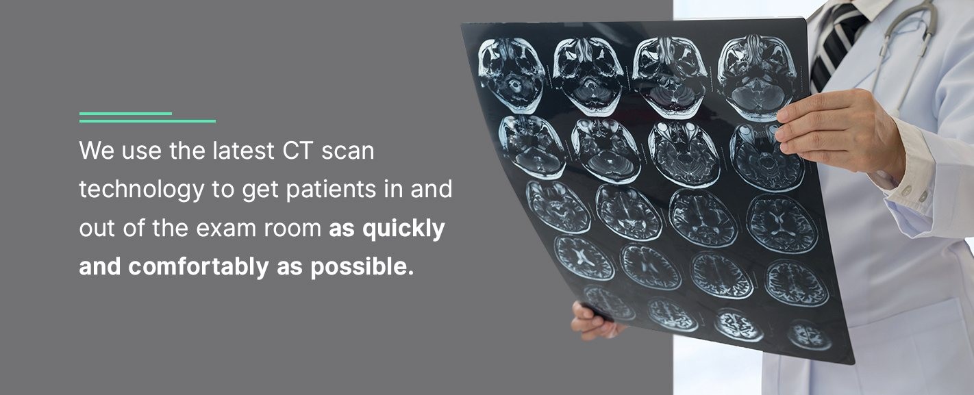
While CT scans are generally painless and straightforward, medical facilities like Health Images prioritize patient comfort and efficiency by employing the latest CT technology to expedite the process.
Understanding PET Scans: Revealing Body Function at a Cellular Level
A Positron Emission Tomography (PET) scan is a nuclear medicine imaging technique. It utilizes a minute amount of radioactive material, known as a radiotracer, along with a specialized camera and computer, to visualize the body’s internal workings. Unlike CT scans that primarily show structure, PET scans reveal how tissues and organs are functioning at a cellular level.
PET scans are most frequently employed in cancer detection and monitoring. They are also valuable in assessing brain and heart function. By highlighting areas of increased metabolic activity, PET scans can detect diseases like cancer in their earliest stages, often before structural changes are visible on CT scans.
How PET Scans Work: Tracers and Metabolic Activity
In a PET scan procedure, a radiotracer is injected into the patient, usually intravenously. This tracer, a radioactive substance attached to a biological molecule like glucose, travels through the body and is absorbed by tissues and organs. The patient then waits for approximately an hour to allow the tracer to distribute and be absorbed. Because cancerous cells typically have a higher metabolic rate, they absorb more of the glucose-based tracer than healthy cells.
After the waiting period, the patient lies on a table that moves into the PET scanner. The scanner detects the radioactive emissions from the tracer and converts this information into three-dimensional images displaying metabolic activity. Hybrid PET/CT scanners are also common, combining the functional information of a PET scan with the anatomical detail of a CT scan in a single imaging session.
PET Scan vs. CT Scan: Key Differences Unveiled
Both PET and CT scans are invaluable diagnostic tools providing clear and accurate images of the body. While they share similarities in their non-invasive nature and application in diagnosing conditions like cancer, the answer to “is a pet scan and ct scan the same?” remains no, due to fundamental differences in their focus and methodology.
Distinguishing Factors Between CT and PET Scans
The primary distinction lies in what each scan visualizes. A CT scan provides a detailed anatomical picture of organs, bones, and tissues, capturing a static image of the body’s structure. Conversely, a PET scan reveals physiological activity at the cellular level, showing how tissues are functioning. Further key differences include:
- Material Usage: CT scans use X-rays that pass through the body to create images. PET scans utilize a radioactive tracer that emits energy, which is then detected by a specialized camera.
- Procedure Duration: CT scans are generally quick, often completed within minutes, making them ideal for emergency situations requiring rapid diagnosis. PET scans are more time-consuming, potentially taking from 20 minutes to several hours depending on the specific scan and patient condition, sometimes even spread over several days.
- Radiation Persistence: No radiation remains in the body after a CT scan. However, a small amount of radiation from the radiotracer may remain in the body for a short period following a PET scan. The radiotracer naturally decays and is eliminated from the body.
- Cancer Detection Timing: PET scans are often capable of detecting cancer earlier than other imaging techniques. By revealing molecular activity, PET scans can identify diseases in their earliest stages, sometimes before structural changes become apparent on CT scans. CT scans typically detect cancer or other issues once they have caused structural changes in tissues or organs.
Shared Characteristics of PET and CT Scans
Despite their differences, PET and CT scans also share several important characteristics:
- Both are typically outpatient procedures, meaning they don’t require hospital admission.
- Both are valuable tools in cancer diagnosis and staging.
- Both are considered accurate, painless, and non-invasive procedures.
- Both can reduce or eliminate the need for exploratory surgery by providing detailed internal images.
Ultimately, both CT and PET scans are crucial medical imaging tests that assist doctors in diagnosing the underlying causes of a patient’s symptoms, leading to effective treatment plans and improved health outcomes.
Your Imaging Appointment: What to Expect
Whether your physician has recommended a CT scan or a PET scan, it’s normal to feel some apprehension. Any medical imaging procedure can evoke anxiety. At Health Images, our dedicated and compassionate team of technologists is committed to making your experience as comfortable and stress-free as possible.
We are equipped with advanced technology and a caring staff to meet your imaging needs with expertise and empathy. To discover more about our services or to schedule your appointment, please contact a Health Images center today.
Call To Schedule Your Appointment
Sources
- https://www.independentimaging.com/ct-scans-vs-pet-scans/
- https://www.ctoam.com/services/testing/imaging/pet-ct-scan/pet-ct-scan-vs-ct/
- https://medlineplus.gov/ctscans.html
- https://www.healthimages.com/services/ct-scans/
- https://www.radiologyinfo.org/en/info.cfm?pg=abdominct
- https://www.mayoclinic.org/tests-procedures/ct-scan/about/pac-20393675
- https://www.radiologyinfo.org/en/info.cfm?pg=pet
- https://www.radiologyinfo.org/en/info.cfm?pg=gennuclear
- https://medlineplus.gov/ency/article/003827.htm
- https://www.mayo.edu/research/documents/radiation-exposure-during-imaging-examspdf/DOC-10027821
- https://my.clevelandclinic.org/health/diagnostics/10123-pet-scan
- https://www.healthimages.com/locations/
- https://www.health.harvard.edu/cancer/radiation-risk-from-medical-imaging
- https://www.healthimages.com/about-us/