Understanding your PET scan results can feel overwhelming, but PETS.EDU.VN is here to help you navigate the medical jargon and complex terminology. This guide clarifies common terms, acronyms, and values, empowering you to understand your report better. PET imaging, nuclear medicine, and diagnostic scans are the keys to a better understanding.
1. What is a PET Scan?
PET stands for Positron Emission Tomography. It’s a nuclear medicine procedure that uses a radioactive substance (radiotracer) to create images of your organs and tissues. The radiotracer emits positrons, which are detected by special cameras as you move through the scanner. These recordings are then compiled into colorful images for interpretation.
PET scans are often used with other imaging tests like CT scans or MRIs to assess both the function and structure of organs and tissues. This combination provides a more complete picture of your health. According to the Society of Nuclear Medicine and Molecular Imaging (SNMMI), PET scans are invaluable for diagnosing and monitoring various conditions, including cancer, heart disease, and neurological disorders.
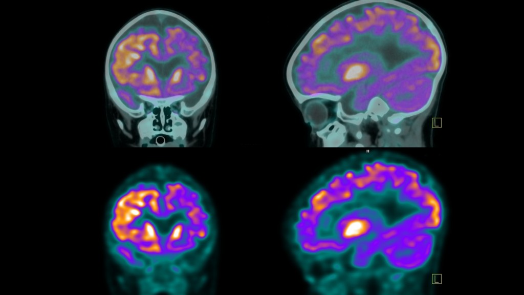
2. Understanding Imaging Planes: Axial, Sagittal, and Coronal
PET scanners capture images along three different planes, providing a comprehensive view of your body’s internal structures. The radiologist can see a 360° view.
- Axial: Also called the transverse view, this is a horizontal view that creates images of the top and bottom halves of your body.
- Sagittal: The side view, providing lateral images of the left and right halves of the body.
- Coronal: The face-on view, creating images of the front and back halves of your body.
Understanding these planes helps visualize the location and extent of any abnormalities detected during the scan. The American College of Radiology (ACR) emphasizes the importance of multiplanar imaging for accurate diagnosis.
3. The Role of Radiotracers in PET Scans
The radiotracer is a radioactive substance used in PET scans. It’s injected, inhaled, or ingested and travels through your body, accumulating in cells that require a lot of energy. A common radiotracer is Fluorodeoxyglucose (FDG). Your PET scan report will detail the radiotracer used, the amount administered, the administration site, and the method used.
The choice of radiotracer depends on the specific area or condition being investigated. For example, different radiotracers are used to detect tumors, assess brain function, or evaluate heart health.
4. Deciphering FDG and FDG Uptake in PET Scan Results
FDG is an abbreviation for Fluorodeoxyglucose, a widely used radiotracer that helps visualize how your body’s cells metabolize glucose. Understanding FDG and its role in PET scans is key to interpreting your imaging results. Terms like “FDG uptake” or “FDG activity” refer to how cells absorb and use this tracer, providing insight into your metabolic health.
PETS.EDU.VN provides in-depth explanations of these terms, ensuring you’re well-informed about your scan results. We break down the science in an accessible way, empowering you to discuss your health with confidence.
4.1. What is FDG Uptake?
FDG uptake describes how much radiotracer was ‘taken up’ by cells. Different cell types have unique metabolic needs, so FDG will cluster at varying concentrations in different areas of the body. FDG activity refers to how vigorously the body’s tissues are utilizing glucose, reflected by how much FDG is absorbed. Higher FDG activity usually indicates higher metabolic activity. Simply put, FDG uptake is a measurement of how much FDG is in the tissues, whereas FDG activity indicates the metabolic qualities that cause the absorption of FDG.
The amount of FDG uptake can indicate various conditions, from normal tissue function to cancerous activity. Interpreting these results requires expertise, and your doctor will consider various factors to determine the significance of the findings.
4.2. Understanding “No Uptake” in PET Scans
No uptake means the FDG isn’t being absorbed. Whether this is a positive or problematic result depends on the baselines of the tissue being studied. For some areas, low metabolic activity is normal; in others, it is a sign of an abnormality. Your doctor will weigh this result against specific tissue types, your overall medical condition, and if this is normal for your baseline. Possible indications:
- Tumor or growth is not active: For areas that previously had growths or tumors, no uptake could indicate that the growth is no longer as metabolically active. This might suggest that the tissue is necrotic (dying), but it could also mean that medical treatments have been successful, and the tumor or growth is no longer growing or active. Your doctor will likely confirm the results with additional testing.
- Normal tissue: Certain tissues and areas of the body are expected to have no FDG uptake or low metabolic activity. In those circumstances, this is considered a healthy result.
- Detection problems: If the scanning machine isn’t sensitive enough or is having technical issues, it may not be able to detect the FDG uptake.
- Not currently inflamed/infected: If the PET scan was to assess an area of the body with infection or inflammation, no FDG uptake could suggest that the problem has been resolved or is not currently active.
4.3. Interpreting Normal FDG Uptake in PET Scan Reports
Areas of your body, such as the spleen, liver, and brain, tend to have higher FDG uptake because they have higher glucose needs and are therefore more metabolically active. Most tissues have established medical baselines for expected FDG absorption, which vary depending on tissue type and location within the body. Normal uptake results suggest that your tissues are functioning within expected metabolic parameters.
Understanding these normal variations is crucial for differentiating healthy tissue from potentially problematic areas. PETS.EDU.VN provides detailed information on typical FDG uptake levels in different organs and tissues.
4.4. The Significance of Mild or Low FDG Uptake
Low or mild FDG uptake can be normal for less active tissues or those high in fat. In some reports, it may also be referred to as low-level or low-grade FDG uptake. However, in areas expected to be more metabolically active, it may indicate the need for closer inspection. Possible explanations for low FDG uptake:
- Less active or non-viable tissue: This could suggest that previously active areas are now necrotic. It might also indicate that a tumor, inflammation, or disease is less active or no longer active, which could be a positive sign of successful treatment. Your doctor will likely perform additional tests to clarify the situation.
- Possible tumor detection: Certain tumors or other conditions may naturally exhibit lower FDG uptake.
- Technical limitations or issues with the scanning equipment: Sometimes, the scanning equipment itself may have sensitivity issues or be malfunctioning, affecting the results.
4.5. What Increased FDG Uptake Really Means
“Increased FDG uptake” or “Intense FDG uptake” on a PET scan means that cells in a certain area of the body are absorbing more of the radiotracer FDG than surrounding tissues. This higher uptake typically appears as brighter or more intense spots on the scan. Increased FDG uptake can indicate:
- Cancer: Cancer cells are usually more active and consume more glucose, resulting in higher FDG uptake.
- Inflammation or Infection: Areas with active inflammation or infection show increased uptake due to heightened immune activity.
- Tissue Healing: Following surgery, injury, or radiation therapy, healing tissues may exhibit increased FDG uptake.
- Benign Conditions: Some non-cancerous growths or active thyroid nodules can also show higher FDG uptake.
While increased FDG uptake often raises concerns for cancer, it can also be due to benign or non-malignant processes. Proper interpretation by a radiologist, considering the clinical context and additional tests, is essential for accurate diagnosis.
4.6. Deciphering Abnormal FDG Uptake in PET Scans
This occurs when glucose absorption is at an irregular level for the tissue being assessed. This can mean either less than expected FDG absorption or higher than expected absorption. For higher FDG uptake, additional testing will typically be conducted to determine the cause. Possible indications for higher FDG uptake levels include:
- Possible cancer: Many types of cancer cells have higher metabolic activity, which leads to a higher FDG absorption rate.
- Infection or inflammation: Increased metabolic activity of immune cells responding to inflammation or infection can also result in higher FDG uptake.
5. SUV: Understanding Standard Uptake Value
SUV is a medical abbreviation for the term Standard Uptake Value, a ratio that defines the activity of the radiotracer (such as FDG) in a specific area of a PET scan image at a specific point in time. It is also known as the dose uptake ratio. A higher SUV may indicate increased metabolic activity, which could be due to various factors, including inflammation, infection, or cancerous growths. Conversely, a lower SUV could indicate less metabolic activity. Generally speaking, metabolic activity is considered:
- “Low intensity” at <5 SUV
- “Moderate” at 5-10 SUV
- “Intense” at 10-15 SUV
- “Very intense” at >15 SUV
The SUV value is helpful for interpretation purposes, especially when comparing multiple PET or other scans over time. The increase or decrease in SUV can give the radiologist a clear understanding of how conditions or treatments are progressing.
The National Institutes of Health (NIH) provides detailed information on SUV calculations and their clinical applications.
6. Physiological Uptake: What It Means for Your PET Scan
The term “physiological uptake” can be confusing, as it’s conceptually similar to FDG uptake. The difference is that physiological uptake is based on the typical, expected absorption of any radiotracer throughout the body. It doesn’t have to be FDG specifically, though PET scans generally use FDG as their radiotracer of choice. The key point to understand is that different organs and tissues have different standardized expectations for radiotracer uptake because metabolic activity differs by organ or tissue type.
Knowing the standard benchmarks for physiological uptake allows physicians to determine if tissues and organs are behaving in a healthy, expected manner. Abnormalities or atypical reactions to radiotracer in these areas may suggest disease, growths, or other health conditions.
6.1. Does Physiological Uptake Mean Cancer?
No, physiological uptake does not mean cancer. Physiological uptake refers to the normal absorption or accumulation of a substance, such as a contrast agent or radiotracer, in the body’s tissues during imaging studies like PET scans. This uptake occurs in organs and tissues that naturally use or process the substance, such as the brain, heart, liver, or kidneys.
While increased uptake in certain areas can sometimes indicate cancer or other abnormalities, physiological uptake is considered a normal finding and not indicative of disease. It’s important to differentiate between physiological and abnormal uptake when interpreting imaging results, and this is typically done by a radiologist or nuclear medicine specialist.
6.2. Understanding Physiological Activity in the Liver
“Physiologic activity in the liver” refers to normal metabolic activity within the liver. This is expected because the liver is a metabolically active organ that plays a key role in various bodily functions, including glucose metabolism, detoxification, and protein synthesis. General findings:
- Normal Finding: The liver naturally absorbs the radiotracer (FDG) due to its high metabolic activity. This is seen as a uniform or mildly increased uptake on the scan.
- Non-Specific: Physiologic activity does not indicate any pathology. It simply reflects the liver’s normal functioning.
- Variations: Levels of uptake can vary slightly based on factors like diet, blood sugar levels, and liver health but are generally within a normal range.
Physiologic activity in the liver on a PET scan is a standard finding and is not associated with any disease.
6.3. Physiological Activity in Kidneys and Bladder: What to Expect
Physiologic uptake in the kidneys and bladder refers to the normal absorption and accumulation of the radiotracer used during the scan.
- Kidneys: After the radiotracer is injected into the bloodstream, it is filtered out by the kidneys as part of the body’s natural process of removing waste. This filtering process results in the normal, or physiologic, uptake of the radiotracer in the kidneys, which will show up on the PET scan.
- Bladder: As the kidneys filter the radiotracer, it is excreted into the urine and accumulates in the bladder. The bladder then shows physiologic uptake as it stores this radiotracer, which is also visible on the PET scan.
This physiologic uptake is expected and indicates that the kidneys and bladder are functioning properly in filtering and excreting the radiotracer. It is not a sign of disease but rather a normal part of the body’s response during the PET scan.
7. Metabolic Activity: A Key Indicator in PET Scans
Metabolic activity on a PET scan refers to how actively cells in the body are using glucose or other metabolic substrates. PET scans detect this activity by using a radiotracer, like FDG, which highlights areas with increased metabolic uptake, helping to identify abnormal or potentially diseased tissue.
PETS.EDU.VN explains how metabolic activity is measured and interpreted, providing a clearer understanding of your scan results. We also offer resources for further research and consultation with medical professionals.
7.1. Interpreting “No Metabolic Activity” in PET Scan Results
No metabolic activity can have various implications on a PET scan. In certain areas of the body, this may be expected if the tissues don’t normally react to glucose or metabolic factors, which is a normal result. However, if this result is seen as an abnormality, it could suggest the following possibilities:
- Damaged Tissue: The tissue could be necrotic or damaged to the extent that it no longer metabolizes FDG effectively.
- Possible Blockages: There may be reduced blood flow or other blockages, such as those resulting from heart attacks or strokes, affecting the area.
- Possible Successful Treatment: In the context of a tumor or growth, a lack of metabolic activity could indicate that the abnormality is no longer active, suggesting a positive response to treatment and potential improvement in health.
7.2. Low-Grade or Mild Metabolic Activity: What Does It Mean?
Low-grade metabolic activity (also called mild metabolic activity) means that less radiotracer is being absorbed. For certain tissues and organs, this may be a normal result due to inherently lower metabolic activity. However, when low-grade metabolic activity is observed as an abnormality, it can be indicative of several conditions:
- Early Stages of Infections or Disease: This might represent the initial phase of conditions such as tumors or infections, where metabolic activity has not yet increased and remains low-grade.
- Scar Tissue: Fibrous tissues resulting from previous surgeries or injuries generally have lower metabolic demands compared to healthy or diseased tissues.
- Aging: Metabolic rate naturally declines with age, so low-grade metabolic activity in older adults can be a normal and expected finding.
- Metabolic Disorders: Certain metabolic disorders can impact metabolic activity and radiotracer uptake. Examples include diabetes, metabolic syndrome, enzyme deficiencies, hypothyroidism and similar conditions.
- Beginning of Successful Treatment/Healing: As tumors, inflammation, or diseases respond to treatment, a reduction in metabolic activity may indicate that healing or treatment efficacy is occurring.
7.3. Increased Metabolic Activity or Hypermetabolic: What Does It Indicate?
Increased metabolic activity, also called hypermetabolic, often indicates that cells are more active than normal, which can be a sign of inflammation, infection, or cancer, as these cells tend to consume more glucose.
“Increased metabolic activity” or “hypermetabolic” on a PET scan indicates areas where cells are more active than normal and tend to consume more glucose. These areas show an increased radiotracer uptake and appear as brighter spots on the scan.
Increased metabolic activity may indicate:
- Cancerous Tumors: Rapidly dividing cancer cells consume more glucose.
- Inflammation and Infection: Active immune responses increase cellular activity.
- Healing and Tissue Repair: Regenerating tissues post-injury or surgery show temporary increases.
- Benign Conditions: Some non-cancerous conditions, such as benign tumors, may also appear hypermetabolic.
While increased metabolic activity can signal serious conditions, it is not always indicative of cancer. A radiologist and doctor will interpret these results based on your medical history and further testing.
8. Deauville Score: A Key Metric in Lymphoma Treatment
The Deauville score or scale (DS) is an internationally recommended standard for reporting FDG uptake in treatment trials for Hodgkin’s and non-Hodgkin’s lymphomas. Like the SUV, it measures FDG uptake, but the Deauville score is a visual interpretation that compares uptake in affected areas to uptake in the liver and mediastinum (the space between the lungs containing the esophagus, heart, large blood vessels, and trachea).
The Deauville score runs from 1 to 5:
- No uptake
- Slight uptake, equal to or below uptake in the mediastinum
- Uptake above the mediastinum but below the liver
- Uptake slightly or moderately above the liver
- Noticeably increased uptake compared to the liver
When it comes to the Deauville score, a lower number is better. 1 and 2 are both considered complete responses. 3 is adequate, while 4 and 5 are considered inadequate.
Understanding the Deauville score helps patients and doctors assess the effectiveness of lymphoma treatment and make informed decisions about ongoing care.
9. Unremarkable: A Positive Finding in Your PET Scan Report
In medical terminology, “unremarkable” is a good thing: it means your PET scan reports no abnormal findings. Your PET scan report is one place you actually want to be found unremarkable.
10. Navigating Your Healthcare Journey with PETS.EDU.VN
PETS.EDU.VN is committed to providing you with the knowledge and resources you need to understand your PET scan results. Our website offers comprehensive information, expert insights, and practical advice to help you navigate your healthcare journey with confidence. We understand that waiting for results can be stressful, and we strive to empower you with the information you need to ask informed questions and make informed decisions.
11. Common Questions About PET Scan Results (FAQ)
- Q1: What is the purpose of a PET scan?
- A1: PET scans are used to detect diseases like cancer, heart problems, and brain disorders by imaging the body’s metabolic processes.
- Q2: How should I prepare for a PET scan?
- A2: Preparation typically involves fasting for a few hours before the scan and avoiding strenuous activity. Your doctor will provide specific instructions.
- Q3: Are there any risks associated with PET scans?
- A3: PET scans involve exposure to small amounts of radiation. Risks are generally low, but you should discuss any concerns with your doctor.
- Q4: How long does a PET scan take?
- A4: The scan duration varies, but it usually takes between 30 minutes to an hour.
- Q5: When will I receive my PET scan results?
- A5: Results are usually available within a few days. Your doctor will schedule a follow-up appointment to discuss the findings.
- Q6: Can I access my PET scan images online?
- A6: Many healthcare providers offer online access to medical images and reports. Check with your provider for availability.
- Q7: What if my PET scan shows abnormal results?
- A7: Abnormal results may require further testing or treatment. Your doctor will guide you through the next steps.
- Q8: Can a PET scan detect all types of cancer?
- A8: PET scans are effective for detecting many types of cancer but may not be suitable for all. Other imaging tests may be necessary.
- Q9: Is a PET scan better than a CT scan or MRI?
- A9: Each imaging test has its strengths. PET scans are excellent for detecting metabolic activity, while CT scans and MRIs provide detailed anatomical images.
- Q10: How much does a PET scan cost?
- A10: The cost of a PET scan varies depending on the location and facility. Contact your insurance provider for coverage details.
12. PET Scan Results: New Advances
| Advancement | Description | Benefits |
|---|---|---|
| Improved Radiotracers | Development of new radiotracers that target specific biomarkers, allowing for more precise imaging of diseases. | Enhanced diagnostic accuracy, earlier disease detection, and improved monitoring of treatment response. |
| Advanced Imaging Techniques | Integration of PET with MRI (PET/MRI) and improvements in PET/CT technology, providing higher resolution and more detailed images. | Better anatomical localization of metabolic activity, reduced radiation exposure, and improved diagnostic confidence. |
| Artificial Intelligence (AI) | Use of AI algorithms to analyze PET scan images, assisting radiologists in detecting subtle patterns and anomalies. | Faster and more accurate image analysis, reduced inter-observer variability, and improved efficiency in clinical workflows. |
| Personalized Medicine | Tailoring PET imaging protocols and radiotracer selection based on individual patient characteristics and genetic profiles. | More precise and individualized diagnostic and treatment strategies, leading to better patient outcomes. |
| Quantitative PET | Development of standardized methods for quantifying PET scan results, such as SUV measurements, enabling more objective and reproducible assessments. | Improved monitoring of disease progression and treatment response, facilitating more informed clinical decision-making. |
| Theranostics | Use of PET imaging to identify patients who are likely to benefit from targeted therapies (theranostics), where the same molecule is used for both imaging and treatment. | More effective selection of patients for targeted therapies, leading to better outcomes and reduced unnecessary treatments. |
| Multi-Parametric PET | Combining PET imaging with other modalities, such as genomic or proteomic data, to provide a more comprehensive understanding of disease biology. | Enhanced characterization of tumors and other diseases, enabling more precise and personalized treatment approaches. |
| Clinical Applications | Expanding the clinical applications of PET imaging beyond oncology to include neurology, cardiology, and infectious diseases. | Earlier and more accurate diagnosis of a wider range of diseases, leading to improved patient management and outcomes. |
| Cost-Effectiveness | Development of strategies to reduce the cost of PET imaging, such as optimizing radiotracer production and streamlining imaging protocols. | Increased accessibility of PET imaging, making it more widely available to patients who can benefit from it. |
| Patient Education | Increased efforts to educate patients about the benefits and limitations of PET imaging, ensuring that they are well-informed and engaged in their healthcare decisions. | Improved patient satisfaction and adherence to treatment plans, leading to better outcomes. |
Understanding your PET scan results is crucial for making informed decisions about your health. At PETS.EDU.VN, we’re dedicated to empowering you with the knowledge and resources you need. If you’re seeking more detailed information or personalized guidance, we encourage you to visit our website at pets.edu.vn or contact us at 789 Paw Lane, Petville, CA 91234, United States, or Whatsapp: +1 555-987-6543. Together, we can navigate your healthcare journey with confidence and clarity.