Positron Emission Tomography (PET) scans are an advanced imaging technique increasingly used in veterinary medicine to provide detailed insights into your pet’s health. These scans go beyond traditional imaging by not just showing the structure of organs and tissues, but also how they are functioning at a cellular level. This functional imaging capability is particularly valuable when it comes to detecting and understanding diseases like cancer in pets.
What are PET Scans and How Do They Work for Pets?
Think of a PET scan as a way to see your pet’s body in action. Unlike X-rays or CT scans that primarily show anatomy, PET scans reveal metabolic activity. This is achieved by using a special tracer, often a radiolabeled glucose analogue called FDG (fludeoxyglucose). FDG is similar to sugar, which cells use for energy. When FDG is injected into your pet, it travels through the bloodstream and is absorbed by tissues.
Cells that are more metabolically active, meaning they are working harder and using more energy, will absorb more FDG. Cancer cells are known for their high metabolic rate. Because malignant tumors grow rapidly and require a lot of energy, they tend to accumulate more FDG than normal, healthy tissues or benign tumors. The PET scanner detects the radioactive signals from the FDG, creating images that highlight areas of high metabolic activity within your pet’s body.
Differentiating Benign from Malignant Tumors with PET Scans
The key advantage of PET scans in oncology is their ability to help differentiate between benign and malignant tumors. While other imaging methods can identify masses or abnormal growths, they may not always be able to determine if these growths are cancerous or not. PET scans, by showing the metabolic activity of these growths, offer a significant advantage.
Malignant tumors, due to their rapid growth and increased metabolism, typically show up as “hot spots” on PET scans, indicating high FDG uptake. Benign tumors or inflammatory conditions, which are less metabolically active, usually exhibit lower FDG uptake, or no increased uptake compared to surrounding normal tissues.
This difference in metabolic activity is crucial for diagnosis. For instance, in cases where a pet has a suspicious mass, a PET scan can help veterinarians determine if it’s more likely to be a benign growth, like a lipoma, or a malignant tumor requiring aggressive treatment. It’s important to note that while PET scans are highly effective, they are usually used in conjunction with other diagnostic tools like biopsies for definitive diagnosis.
Benefits of PET Scans in Pet Cancer Diagnosis and Management
Beyond differentiating benign and malignant tumors, PET scans offer several other benefits in managing pet cancer:
-
Early Detection and Staging: PET scans can detect metabolically active tumors even when they are small and not yet visible on other imaging modalities. This early detection can be critical for successful treatment. Furthermore, PET scans are valuable for staging cancer, determining if and where the cancer has spread (metastasis) in the body.
-
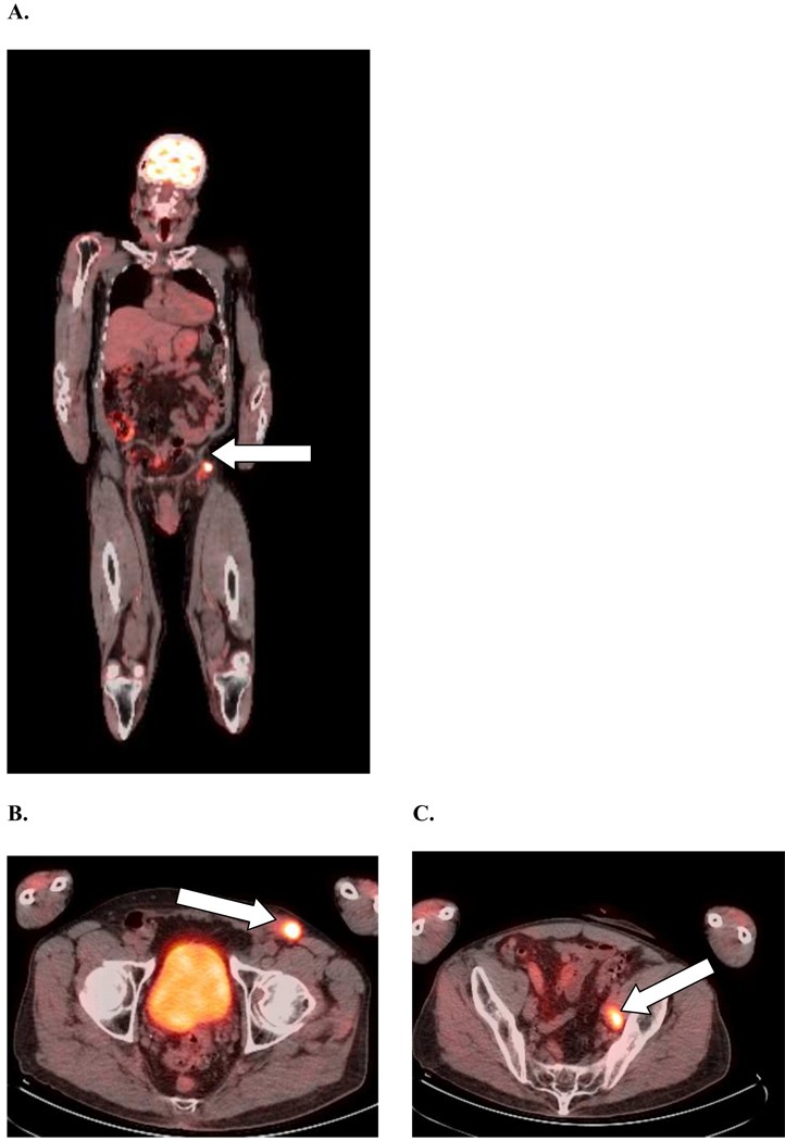
More Accurate Extent of Disease: By highlighting metabolically active areas, PET scans provide a more accurate picture of the extent of the tumor, which is crucial for surgical planning and radiation therapy. As shown in the original human example, a PET-CT scan revealed unexpected bone metastasis, which significantly altered the treatment approach from surgery to systemic therapy. This same principle applies to pets.
- Monitoring Treatment Response and Detecting Recurrence: PET scans can be used to monitor how well a tumor is responding to treatment, such as chemotherapy or radiation therapy. A decrease in FDG uptake can indicate that the treatment is effective. They can also help in detecting recurrence of cancer earlier than other methods by identifying areas of increased metabolic activity that might suggest the cancer has returned.
Considerations and Limitations
While PET scans are a powerful tool, it’s important to understand their limitations. Not all tumors are FDG-avid, meaning some tumors may not show up clearly on a PET scan. Also, inflammation and infection can also cause increased FDG uptake, leading to false positives. Therefore, PET scan results should always be interpreted in conjunction with other clinical findings and diagnostic tests.
Conclusion
PET scans represent a significant advancement in veterinary oncology, offering a unique functional perspective for diagnosing and managing cancer in pets. Their ability to differentiate between benign and malignant tumors, along with their benefits in staging, treatment planning, and monitoring, makes them an invaluable tool for veterinarians striving to provide the best possible care for pets with cancer. If your pet is facing a potential cancer diagnosis, discuss with your veterinarian whether a PET scan could be beneficial in their diagnostic and treatment journey.