Prostate cancer diagnosis and management have been revolutionized by advancements in medical imaging. Among these innovations, the PSMA PET scan stands out as a highly effective method for detecting prostate cancer throughout the body. This article delves into the capabilities of PET scans in prostate cancer detection, focusing on the cutting-edge PSMA PET technology.
What is PSMA PET Imaging for Prostate Cancer?
PSMA PET imaging is a sophisticated diagnostic technique that utilizes positron emission tomography (PET) to visualize prostate cancer cells, regardless of their location in the body. PSMA, or prostate-specific membrane antigen, is a protein that is highly expressed on the surface of prostate cancer cells. This characteristic makes PSMA an excellent target for imaging.
How Do PSMA PET Scans Work and What Are Their Advantages?
PET scans have been employed in cancer detection for many years. However, traditional PET radiotracers were not particularly effective at imaging prostate cancer. The breakthrough came with the development of new radiotracers, such as piflufolastat F-18 (Pylarify), that specifically bind to PSMA on prostate cancer cells.
During a PSMA PET scan, this radiotracer is administered to the patient. It circulates through the body and attaches to prostate cancer cells, which then “light up” on the PET scan images. This allows doctors to pinpoint the precise location of prostate cancer, even in very small tumors or distant metastases.
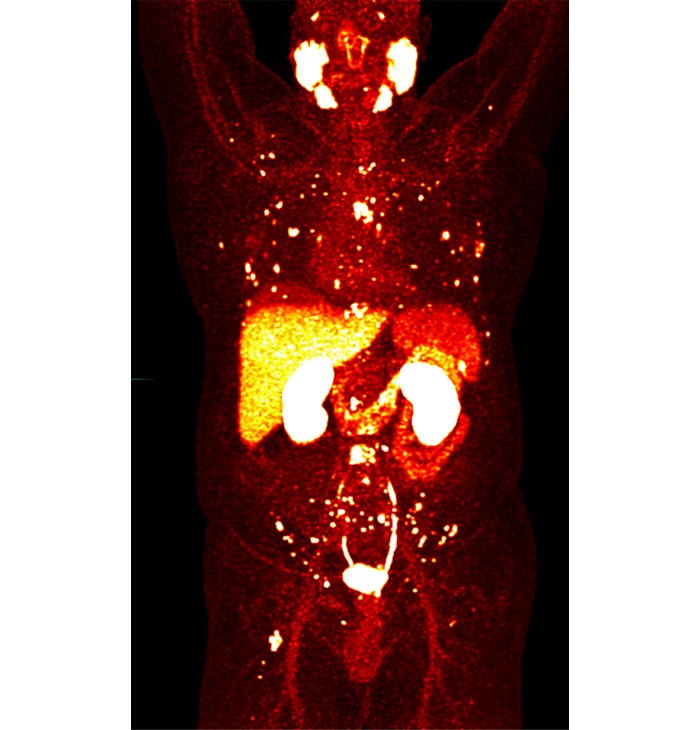
Alt text: Full body PSMA PET scan demonstrating areas of prostate cancer indicated by bright spots, highlighting the scan’s sensitivity in detecting tumors.
The primary benefit of PSMA PET scans is their superior ability to visualize prostate cancer compared to older imaging methods. This enhanced visualization leads to more informed treatment decisions and improved monitoring of a patient’s response to therapy. Knowing the precise extent and location of the cancer is crucial for effective management.
PSMA PET Scans vs. Traditional Prostate Cancer Imaging (CT, MRI, Bone Scans)
Conventional imaging techniques like CT scans, MRI, and bone scans have limitations in detecting small prostate tumors or early-stage disease spread. PSMA PET scans operate at a molecular level, directly targeting the tumor cells. This molecular targeting provides a significant advantage in detecting prostate cancer earlier and more accurately.
PSMA PET scans excel in identifying very small tumors that might be missed by CT, MRI, or bone scans. This is because PSMA PET detects the biological activity of the cancer cells, whereas CT and MRI primarily show anatomical changes which may not be evident in early or microscopic disease.
Alt text: Side-by-side comparison of a CT scan and a PSMA PET scan, illustrating the PSMA PET scan’s ability to detect prostate cancer in a small lymph node missed by the CT scan, showcasing PSMA PET’s enhanced sensitivity.
The image above illustrates this point. A CT scan failed to detect prostate cancer in a small lymph node, whereas a PSMA PET scan clearly identified the cancerous cells. This demonstrates the superior sensitivity of PSMA PET imaging.
Who is the Right Candidate for PSMA PET Imaging?
PSMA PET scans are particularly beneficial for patients in specific clinical scenarios. They are approved for individuals whose prostate cancer is suspected to have spread beyond the prostate and is potentially treatable with radiation, surgery, or systemic therapies. Furthermore, PSMA PET is valuable for patients with suspected prostate cancer recurrence, indicated by a rising PSA level after initial treatment. In essence, if accurate staging and localization of prostate cancer are needed to guide treatment decisions, PSMA PET scanning is a strong consideration.
What to Expect During a PSMA PET Scan Procedure
Undergoing a PSMA PET scan is a straightforward process that typically requires minimal preparation.
- Radiotracer Injection: Upon arrival at the clinic, patients change into a hospital gown and receive an intravenous injection of the piflufolastat F-18 radiotracer.
- Waiting Period: There is a waiting period of approximately one hour to allow the radiotracer to distribute throughout the body and be absorbed by any prostate cancer cells.
- PET Scan: The patient is then positioned in the PET scanner and instructed to lie still for about 20 minutes while the machine acquires images of the body.
- Procedure Completion: Once the scan is finished, the patient can usually leave the clinic without restrictions.
- Image Evaluation: Nuclear medicine physicians analyze the scans, typically on the same day, and generate a report. This report and the PET images are then reviewed by the patient’s cancer doctor to guide treatment planning.
Is the PSMA PET Scan Radiotracer FDA Approved and Safe?
Yes, the radiotracer piflufolastat (Pylarify) used in PSMA PET scans is approved by the FDA and considered safe. While side effects are rare and generally temporary, some patients may experience headache or a temporary alteration in taste.
As a radionuclide dye, Pylarify contains a small amount of radiation, comparable to that of a CT scan. Importantly, the radioactive element is eliminated from the body within a few days, minimizing long-term radiation exposure.
What Happens if a PSMA PET Scan Detects Tumors?
The findings of a PSMA PET scan are crucial for determining the most appropriate treatment strategy. If the scan reveals tumors, the treatment approach will be tailored to the individual patient’s specific situation. Treatment options may include targeted therapy directed at the identified cancer sites, systemic therapy to treat cancer throughout the body, or a combination of both.
In some cases, patients may be candidates for innovative theranostic treatments like lutetium-177 PSMA therapy (Pluvicto). This therapy, related to PSMA PET imaging, also targets the PSMA molecule but uses a more potent form of radiation to destroy cancer cells.
Why Choose PSMA PET Imaging?
PSMA PET imaging represents a significant advancement in prostate cancer care. Its ability to detect prostate cancer with greater accuracy and at earlier stages compared to traditional imaging methods makes it an invaluable tool for diagnosis, staging, treatment planning, and monitoring. For patients and clinicians alike, PSMA PET offers enhanced clarity and confidence in managing prostate cancer.
Accessing PSMA PET Scans
To undergo a PSMA PET scan, a physician’s order is necessary. Individuals interested in PSMA PET scans should discuss this option with their healthcare provider to determine if it is appropriate for their specific medical situation and to navigate insurance coverage.
References: