Can A Pet Scan Detect Kidney Stones? Discover the truth about using PET scans for kidney stone detection with PETS.EDU.VN’s expert insights. Explore alternative imaging techniques and prevention strategies to maintain optimal kidney health.
1. Understanding Kidney Stones: Formation, Types, and Risk Factors
Kidney stones are solid masses made of minerals and salts that form inside your kidneys. These stones can range in size from a grain of sand to a pearl and can cause significant pain and discomfort. According to the National Institute of Diabetes and Digestive and Kidney Diseases (NIDDK), approximately 11% of men and 6% of women in the United States will develop kidney stones at some point in their lives. Understanding the formation, types, and risk factors associated with kidney stones is crucial for prevention and effective management. PETS.EDU.VN offers in-depth resources to help you learn more about kidney stones and how to keep your kidneys healthy.
1.1 How Kidney Stones Form
Kidney stones form when certain substances in the urine, such as calcium, oxalate, uric acid, and cystine, become highly concentrated. When these substances don’t dissolve completely, they can crystallize and stick together, forming stones. Several factors can contribute to this process, including dehydration, diet, medical conditions, and genetics. Maintaining adequate hydration is one of the most important steps you can take to prevent kidney stone formation.
1.2 Common Types of Kidney Stones
There are four main types of kidney stones, each with different compositions and causes:
-
Calcium Stones: The most common type, calcium stones, are usually made of calcium oxalate but can also consist of calcium phosphate. Oxalate is found in many foods, and high doses of vitamin D or certain metabolic disorders can increase the concentration of calcium or oxalate in the urine, leading to stone formation.
-
Struvite Stones: These stones are typically caused by urinary tract infections (UTIs). Bacteria can produce ammonia in the urine, which raises the pH and promotes the formation of struvite stones. These stones can grow quickly and become quite large, often requiring surgical removal.
-
Uric Acid Stones: Uric acid stones form when there is too much uric acid in the urine. This can occur in people who eat a high-protein diet, have gout, or undergo chemotherapy. Certain genetic factors and metabolic disorders can also increase uric acid levels.
-
Cystine Stones: Cystine stones are rare and occur in people with cystinuria, a genetic disorder that causes the kidneys to excrete too much of the amino acid cystine. This excess cystine can lead to the formation of stones.
Knowing the type of kidney stone you have is essential for determining the best course of treatment and implementing strategies to prevent future stone formation. PETS.EDU.VN provides detailed information on each type of kidney stone, including specific dietary recommendations and medical interventions.
1.3 Risk Factors for Developing Kidney Stones
Several factors can increase your risk of developing kidney stones. Being aware of these risk factors can help you take proactive steps to protect your kidney health:
-
Dehydration: Not drinking enough water can lead to more concentrated urine, making it easier for minerals and salts to crystallize and form stones.
-
Diet: A diet high in protein, sodium, and oxalate can increase the risk of kidney stones. Limiting these substances in your diet can help prevent stone formation.
-
Obesity: Being overweight or obese is associated with a higher risk of kidney stones. Obesity can alter the acidity of urine and increase the excretion of calcium and uric acid.
-
Medical Conditions: Certain medical conditions, such as hyperparathyroidism, gout, Crohn’s disease, and renal tubular acidosis, can increase the risk of kidney stones.
-
Family History: If you have a family history of kidney stones, you are more likely to develop them yourself.
-
Certain Medications: Some medications, such as diuretics and certain antacids, can increase the risk of kidney stones.
By understanding these risk factors, you can make informed lifestyle choices and work with your healthcare provider to manage your risk of developing kidney stones. Remember, PETS.EDU.VN is here to provide you with the resources and information you need to stay healthy.
 Understanding Kidney Stones
Alt text: Medical image showing different types of kidney stones and their formation.
2. The Role of PET Scans in Medical Imaging
Positron Emission Tomography (PET) scans are a powerful imaging technique used in medicine to visualize the body’s metabolic processes. Unlike other imaging methods that primarily show the structure of organs and tissues, PET scans provide information about how well these tissues and organs are functioning. PET scans involve injecting a small amount of radioactive tracer, called a radiotracer, into the patient. The radiotracer emits positrons, which interact with electrons in the body, producing gamma rays that are detected by the PET scanner. The scanner then creates detailed, three-dimensional images of the body, highlighting areas of high metabolic activity.
2.1 How PET Scans Work
The radiotracer used in a PET scan is typically a glucose analog, such as fluorodeoxyglucose (FDG), which is similar to glucose but contains a radioactive fluorine atom. Because cancer cells often have a higher metabolic rate than normal cells, they tend to absorb more FDG, making them visible on the PET scan. This allows doctors to detect cancerous tumors and assess their activity.
During a PET scan, the patient lies on a table that slides into a large, donut-shaped scanner. The scan usually takes between 30 minutes to an hour, depending on the area of the body being examined. The amount of radiation exposure from a PET scan is generally low and considered safe for most patients.
2.2 Common Applications of PET Scans
PET scans are primarily used in oncology (cancer diagnosis and treatment), neurology (brain disorders), and cardiology (heart conditions).
-
Oncology: PET scans are used to detect and stage cancer, assess the effectiveness of cancer treatments, and monitor for cancer recurrence. They can help differentiate between benign and malignant tumors and guide treatment decisions.
-
Neurology: PET scans can help diagnose and monitor brain disorders such as Alzheimer’s disease, Parkinson’s disease, and epilepsy. They can also be used to evaluate brain function after a stroke or traumatic brain injury.
-
Cardiology: PET scans can help assess blood flow to the heart and identify areas of damaged or ischemic tissue. They can also be used to evaluate the effectiveness of cardiac treatments, such as bypass surgery and angioplasty.
2.3 Advantages and Limitations of PET Scans
PET scans offer several advantages over other imaging techniques, including their ability to provide information about metabolic activity and detect diseases at an early stage. However, they also have some limitations:
-
High Cost: PET scans are relatively expensive compared to other imaging methods, such as X-rays and CT scans.
-
Radiation Exposure: Although the amount of radiation exposure from a PET scan is low, it is still a concern for some patients, especially pregnant women and children.
-
Limited Availability: PET scanners are not available in all hospitals and clinics, which can limit access to this imaging technique.
-
Image Resolution: PET scans have lower spatial resolution compared to CT scans and MRI, which can make it difficult to visualize small anatomical details.
Despite these limitations, PET scans remain a valuable tool in modern medicine, providing unique insights into the body’s metabolic processes and helping doctors diagnose and treat a wide range of diseases. For more information on medical imaging and diagnostic techniques, visit PETS.EDU.VN.
3. Can a PET Scan Detect Kidney Stones? The Answer Explained
While PET scans are valuable for detecting metabolic activity related to cancer, neurological disorders, and heart conditions, they are generally not the primary imaging modality for detecting kidney stones. The reason lies in what PET scans are designed to visualize: metabolic processes rather than physical structures. Kidney stones are dense, mineral-based structures, and PET scans are not optimized to detect these types of objects.
3.1 Why PET Scans Are Not Suitable for Kidney Stone Detection
-
Focus on Metabolic Activity: PET scans rely on the uptake of radiotracers by metabolically active cells. Kidney stones, being non-metabolic, do not absorb these tracers, making them invisible on a PET scan.
-
Poor Spatial Resolution: PET scans have relatively low spatial resolution compared to other imaging techniques like CT scans. This means that small objects, such as kidney stones, may not be clearly visible, even if they were to interact with the radiotracer.
-
Alternative Imaging Modalities: More effective imaging techniques are available for detecting kidney stones, such as CT scans, X-rays, and ultrasounds. These methods are specifically designed to visualize dense structures within the body.
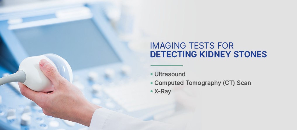
3.2 What Imaging Techniques Are Best for Detecting Kidney Stones?
Several imaging techniques are better suited for detecting kidney stones. These include:
-
Computed Tomography (CT) Scan: CT scans are the gold standard for kidney stone detection. They provide detailed images of the kidneys, ureters, and bladder, allowing doctors to identify even small stones. CT scans are fast, non-invasive, and highly accurate.
-
X-Ray (KUB): A kidney, ureter, and bladder (KUB) X-ray can detect larger kidney stones. However, it is less sensitive than a CT scan and may miss smaller stones.
-
Ultrasound: Ultrasound is a non-invasive imaging technique that uses sound waves to create images of the kidneys. It is often used as the first-line imaging test for pregnant women and children to avoid radiation exposure. However, ultrasound may not be as sensitive as CT scans for detecting small stones.
3.3 When Might a PET Scan Be Used in Conjunction with Kidney Stone Evaluation?
In rare cases, a PET scan might be used in conjunction with kidney stone evaluation if there is suspicion of an underlying metabolic disorder contributing to stone formation. For example, if a patient has recurrent kidney stones and other symptoms suggestive of a metabolic disease, a PET scan might be used to investigate the possibility of an underlying endocrine tumor or other metabolic abnormality. However, this is not a routine application of PET scans in the context of kidney stones.
To summarize, while PET scans are invaluable for certain medical applications, they are not appropriate for detecting kidney stones. Alternative imaging techniques like CT scans, X-rays, and ultrasounds are much more effective for this purpose. For comprehensive information on kidney stone diagnosis and treatment, visit PETS.EDU.VN.
Alt text: Image depicting various kidney stone detection techniques, including CT scan, X-ray, and ultrasound.
4. Alternative Imaging Techniques for Kidney Stone Detection
As established, PET scans are not the go-to method for identifying kidney stones. Instead, doctors rely on other imaging techniques that are better suited for visualizing these mineral deposits. Here’s a closer look at the alternative imaging methods commonly used:
4.1 Computed Tomography (CT) Scan: The Gold Standard
CT scans are widely considered the gold standard for detecting kidney stones due to their high sensitivity and specificity. They provide detailed, cross-sectional images of the kidneys, ureters, and bladder, allowing doctors to identify even small stones with great accuracy.
-
How it works: A CT scan uses X-rays to create detailed images of the body. The patient lies on a table that slides into a donut-shaped scanner, which rotates around the body, capturing images from multiple angles. These images are then processed by a computer to create a three-dimensional view of the kidneys and urinary tract.
-
Advantages:
- High sensitivity and specificity for detecting kidney stones.
- Ability to identify stones of all sizes and compositions.
- Fast and non-invasive procedure.
- Can detect other abnormalities in the kidneys and urinary tract.
-
Disadvantages:
- Involves exposure to radiation.
- May require the use of intravenous contrast dye, which can cause allergic reactions or kidney damage in some patients.
4.2 X-Ray (KUB): A Simple and Quick Option
An X-ray of the kidneys, ureters, and bladder (KUB) is a simple and quick imaging test that can detect larger kidney stones. While not as sensitive as a CT scan, it is often used as the initial imaging test due to its lower cost and radiation exposure.
-
How it works: The patient lies on a table, and an X-ray machine is positioned over the abdomen. X-rays are passed through the body, creating an image of the kidneys, ureters, and bladder on a film or digital detector.
-
Advantages:
- Quick and easy procedure.
- Lower cost compared to CT scans.
- Lower radiation exposure compared to CT scans.
-
Disadvantages:
- Less sensitive than CT scans, especially for small stones.
- Cannot detect all types of kidney stones (e.g., uric acid stones may not be visible).
- Limited ability to visualize other abnormalities in the kidneys and urinary tract.
4.3 Ultrasound: A Radiation-Free Alternative
Ultrasound is a non-invasive imaging technique that uses sound waves to create images of the kidneys. It is often used as the first-line imaging test for pregnant women and children to avoid radiation exposure.
-
How it works: A handheld device called a transducer is placed on the skin over the kidneys. The transducer emits sound waves, which bounce off the kidneys and other structures in the urinary tract. These sound waves are then processed by a computer to create images.
-
Advantages:
- No radiation exposure.
- Non-invasive and painless procedure.
- Relatively low cost.
- Can be performed in real-time.
-
Disadvantages:
- Less sensitive than CT scans, especially for small stones and stones located in the ureters.
- Image quality can be affected by body habitus and bowel gas.
- May not be able to visualize all abnormalities in the kidneys and urinary tract.
4.4 Intravenous Pyelogram (IVP): A Less Common Technique
An intravenous pyelogram (IVP) is an X-ray test that uses contrast dye to visualize the kidneys, ureters, and bladder. While it was once a common imaging test for kidney stones, it has largely been replaced by CT scans due to their higher sensitivity and ability to provide more detailed images.
-
How it works: Contrast dye is injected into a vein in the arm. The dye travels through the bloodstream to the kidneys, where it is filtered and excreted into the urine. X-rays are taken at various intervals to visualize the kidneys, ureters, and bladder as the dye passes through them.
-
Advantages:
- Can provide information about the function of the kidneys and urinary tract.
- Can detect obstructions and other abnormalities.
-
Disadvantages:
- Involves exposure to radiation.
- Requires the use of intravenous contrast dye, which can cause allergic reactions or kidney damage in some patients.
- Less sensitive than CT scans for detecting kidney stones.
- More time-consuming than CT scans and other imaging tests.
In summary, while PET scans are not used for detecting kidney stones, several other imaging techniques are available. CT scans are the gold standard, while X-rays and ultrasounds are often used as initial imaging tests. For more information on kidney stone diagnosis and treatment, visit PETS.EDU.VN.
Alt text: Comparison of CT scan, X-ray, and ultrasound images for kidney stone detection.
5. Symptoms and Diagnosis of Kidney Stones
Kidney stones can cause a range of symptoms, depending on their size and location. Some small stones may pass through the urinary tract without causing any noticeable symptoms, while larger stones can cause severe pain and other complications. Recognizing the symptoms of kidney stones and seeking prompt medical attention is crucial for accurate diagnosis and effective management. PETS.EDU.VN provides valuable resources to help you understand the symptoms, diagnostic process, and treatment options for kidney stones.
5.1 Common Symptoms of Kidney Stones
-
Severe Pain: The most common symptom of kidney stones is severe pain in the side and back, below the ribs. This pain, known as renal colic, can be excruciating and may come in waves. It is caused by the stone blocking the flow of urine and causing the ureter to spasm.
-
Pain Radiating to the Lower Abdomen and Groin: As the stone moves down the ureter, the pain may radiate to the lower abdomen and groin. Men may experience pain in the testicles.
-
Nausea and Vomiting: The intense pain caused by kidney stones can trigger nausea and vomiting.
-
Blood in the Urine (Hematuria): Kidney stones can irritate the lining of the urinary tract, causing blood to appear in the urine. The urine may be pink, red, or brown.
-
Frequent Urination: If a kidney stone is located near the bladder, it can cause a frequent urge to urinate.
-
Painful Urination (Dysuria): Kidney stones can cause pain or a burning sensation during urination.
-
Small Amounts of Urine: The stone may partially or completely block the flow of urine, leading to small amounts of urine being passed.
-
Cloudy or Foul-Smelling Urine: A urinary tract infection (UTI) can sometimes accompany kidney stones, leading to cloudy or foul-smelling urine.
5.2 The Diagnostic Process for Kidney Stones
If you experience symptoms of kidney stones, your doctor will perform a thorough evaluation to confirm the diagnosis and determine the size and location of the stone. The diagnostic process typically involves:
-
Medical History and Physical Exam: Your doctor will ask about your symptoms, medical history, and family history of kidney stones. They will also perform a physical exam to assess your overall health.
-
Urinalysis: A urine sample will be tested for blood, crystals, and signs of infection.
-
Blood Tests: Blood tests may be performed to assess kidney function and check for elevated levels of calcium, uric acid, or other substances that can contribute to kidney stone formation.
-
Imaging Tests: As discussed earlier, imaging tests such as CT scans, X-rays, and ultrasounds are used to visualize the kidneys and urinary tract and identify kidney stones.
5.3 When to Seek Medical Attention
It is important to seek medical attention if you experience any of the following symptoms:
- Severe pain in the side and back
- Pain that is accompanied by nausea, vomiting, fever, or chills
- Blood in the urine
- Inability to pass urine
Prompt medical attention can help prevent complications such as infection, kidney damage, and urinary obstruction. Visit PETS.EDU.VN for more information on when to seek medical care for kidney stones and how to manage your symptoms.
Alt text: Diagram illustrating common symptoms of kidney stones, including pain, nausea, and blood in urine.
6. Treatment Options for Kidney Stones
The treatment for kidney stones depends on the size, location, and composition of the stone, as well as the severity of your symptoms. Small stones may pass on their own with conservative treatment, while larger stones may require more aggressive interventions. PETS.EDU.VN provides comprehensive information on the various treatment options available for kidney stones, helping you make informed decisions about your care.
6.1 Conservative Treatment: Waiting for the Stone to Pass
For small kidney stones that are not causing significant pain or obstruction, conservative treatment may be recommended. This involves:
-
Pain Management: Over-the-counter pain relievers such as ibuprofen or naproxen can help alleviate mild to moderate pain. Your doctor may prescribe stronger pain medications if needed.
-
Increased Fluid Intake: Drinking plenty of water (2-3 liters per day) can help flush the stone out of the urinary tract.
-
Alpha-Blockers: These medications relax the muscles in the ureter, making it easier for the stone to pass.
-
Monitoring: Your doctor will monitor your progress with regular check-ups and imaging tests to ensure that the stone is passing and that there are no complications.
6.2 Medical Procedures for Kidney Stone Removal
If a kidney stone is too large to pass on its own or is causing significant pain or obstruction, medical procedures may be necessary to remove the stone. Common procedures include:
-
Extracorporeal Shock Wave Lithotripsy (ESWL): ESWL uses shock waves to break the stone into smaller pieces that can be passed in the urine. It is a non-invasive procedure that is typically performed on an outpatient basis.
-
Ureteroscopy: Ureteroscopy involves inserting a thin, flexible tube with a camera attached (ureteroscope) into the ureter to locate and remove the stone. The stone may be removed with a basket or broken into smaller pieces with a laser or other device.
-
Percutaneous Nephrolithotomy (PCNL): PCNL is a surgical procedure used to remove large kidney stones. It involves making a small incision in the back and inserting a tube directly into the kidney to remove the stone.
-
Open Surgery: Open surgery is rarely necessary for kidney stone removal but may be required in complex cases.
6.3 Dietary and Lifestyle Modifications to Prevent Future Stones
After a kidney stone has been treated, it is important to take steps to prevent future stones from forming. This may involve:
-
Dietary Changes:
- Increase Fluid Intake: Aim for 2-3 liters of water per day.
- Limit Sodium Intake: High sodium intake can increase calcium excretion in the urine, leading to stone formation.
- Limit Animal Protein: High protein intake can increase uric acid levels in the urine.
- Limit Oxalate-Rich Foods: If you have calcium oxalate stones, limit foods high in oxalate, such as spinach, rhubarb, nuts, and chocolate.
- Maintain Adequate Calcium Intake: Getting enough calcium from your diet can help prevent calcium oxalate stones.
-
Lifestyle Changes:
- Maintain a Healthy Weight: Obesity is associated with a higher risk of kidney stones.
- Exercise Regularly: Regular exercise can help prevent kidney stones.
-
Medications:
- Your doctor may prescribe medications to help prevent specific types of kidney stones from forming. For example, thiazide diuretics can help reduce calcium levels in the urine, while allopurinol can help lower uric acid levels.
By following these treatment options and preventive measures, you can effectively manage kidney stones and reduce your risk of recurrence. For more detailed information and personalized advice, visit PETS.EDU.VN.
Alt text: Overview of treatment options for kidney stones, including conservative management and medical procedures.
7. Prevention Strategies for Kidney Stones
Preventing kidney stones is often more effective than treating them. By adopting certain dietary and lifestyle habits, you can significantly reduce your risk of developing these painful conditions. PETS.EDU.VN provides evidence-based strategies to help you maintain kidney health and prevent kidney stone formation.
7.1 Hydration: The Key to Kidney Health
Staying well-hydrated is one of the most important steps you can take to prevent kidney stones. When you drink enough fluids, your urine becomes less concentrated, making it harder for minerals and salts to crystallize and form stones.
-
How Much Water Should You Drink? Aim for at least 2-3 liters (about 8-12 cups) of water per day. You may need to drink more if you are physically active, live in a hot climate, or have certain medical conditions.
-
What Types of Fluids Are Best? Water is the best choice, but you can also include other fluids such as herbal teas, clear soups, and diluted fruit juices. Avoid sugary drinks, such as sodas and sweetened beverages, as they can increase your risk of kidney stones.
-
How to Stay Hydrated:
- Carry a water bottle with you and refill it throughout the day.
- Drink water before, during, and after exercise.
- Set reminders on your phone to drink water regularly.
- Eat fruits and vegetables with high water content, such as watermelon, cucumbers, and lettuce.
7.2 Dietary Recommendations for Kidney Stone Prevention
Your diet plays a significant role in kidney stone formation. Making certain dietary changes can help reduce your risk of developing stones:
-
Limit Sodium Intake: High sodium intake can increase calcium excretion in the urine, leading to stone formation. Aim for less than 2,300 milligrams of sodium per day.
-
Limit Animal Protein: High protein intake can increase uric acid levels in the urine. Choose lean protein sources and limit your intake of red meat, poultry, and fish.
-
Maintain Adequate Calcium Intake: Getting enough calcium from your diet can help prevent calcium oxalate stones. Aim for 1,000-1,200 milligrams of calcium per day from foods such as dairy products, leafy green vegetables, and fortified foods.
-
Limit Oxalate-Rich Foods: If you have calcium oxalate stones, limit foods high in oxalate, such as spinach, rhubarb, nuts, and chocolate.
-
Increase Citrate Intake: Citrate can help prevent calcium stones from forming by binding to calcium in the urine. Citrus fruits such as lemons, limes, and oranges are good sources of citrate. You can also add lemon or lime juice to your water.
7.3 Lifestyle Modifications for Kidney Stone Prevention
In addition to hydration and dietary changes, certain lifestyle modifications can help prevent kidney stones:
-
Maintain a Healthy Weight: Obesity is associated with a higher risk of kidney stones. Aim for a healthy weight through diet and exercise.
-
Exercise Regularly: Regular exercise can help prevent kidney stones by improving overall health and reducing the risk of obesity.
-
Avoid Excessive Alcohol Consumption: Excessive alcohol consumption can lead to dehydration and increase the risk of kidney stones.
-
Manage Stress: Chronic stress can affect kidney function and increase the risk of kidney stones. Practice stress-reducing techniques such as yoga, meditation, or deep breathing exercises.
By incorporating these prevention strategies into your daily routine, you can significantly reduce your risk of developing kidney stones and maintain optimal kidney health. Visit PETS.EDU.VN for more tips and resources on kidney stone prevention.
Alt text: Illustration depicting lifestyle and dietary tips for preventing kidney stones.
8. Living with Kidney Stones: Management and Support
Living with kidney stones can be challenging, especially if you experience frequent episodes or chronic pain. However, with proper management and support, you can effectively control your symptoms and improve your quality of life. PETS.EDU.VN provides resources and information to help you navigate life with kidney stones, offering practical advice and emotional support.
8.1 Managing Pain and Discomfort
-
Pain Relievers: Over-the-counter pain relievers such as ibuprofen or naproxen can help alleviate mild to moderate pain. Your doctor may prescribe stronger pain medications if needed.
-
Heat Therapy: Applying a warm compress or taking a warm bath can help relax the muscles in the urinary tract and relieve pain.
-
Hydration: Drinking plenty of water can help flush the stone out of the urinary tract and reduce pain.
-
Alpha-Blockers: These medications relax the muscles in the ureter, making it easier for the stone to pass and reducing pain.
8.2 Monitoring and Follow-Up Care
-
Regular Check-Ups: Your doctor will schedule regular check-ups to monitor your kidney function and check for new stones.
-
Imaging Tests: Periodic imaging tests, such as CT scans or ultrasounds, may be performed to assess the size and location of any stones.
-
Urine and Blood Tests: Regular urine and blood tests can help monitor your kidney function and identify any underlying metabolic abnormalities.
8.3 Emotional Support and Coping Strategies
-
Support Groups: Joining a support group can provide you with a sense of community and help you connect with others who understand what you are going through.
-
Counseling: Talking to a therapist or counselor can help you cope with the emotional challenges of living with kidney stones.
-
Stress Management: Practicing stress-reducing techniques such as yoga, meditation, or deep breathing exercises can help improve your overall well-being.
-
Education: Learning as much as you can about kidney stones can help you feel more in control and better prepared to manage your condition. PETS.EDU.VN offers a wealth of information on kidney stones, including prevention strategies, treatment options, and tips for living with kidney stones.
8.4 When to Seek Emergency Care
Seek emergency medical attention if you experience any of the following symptoms:
- Severe pain that is not relieved by pain relievers
- Fever or chills
- Blood in the urine
- Inability to pass urine
- Nausea and vomiting
These symptoms may indicate a serious complication, such as infection or urinary obstruction, which requires prompt medical attention.
By following these management strategies and seeking support when needed, you can effectively live with kidney stones and maintain a good quality of life. Remember, PETS.EDU.VN is here to provide you with the resources and information you need to stay healthy and informed.
Alt text: People supporting each other, representing the emotional support needed for living with kidney stones.
9. Expert Insights and the Future of Kidney Stone Detection
The field of kidney stone detection and treatment is continually evolving, with ongoing research aimed at improving diagnostic accuracy and developing less invasive treatment options. Gaining insights from experts in the field can help you stay informed about the latest advancements and make informed decisions about your care. PETS.EDU.VN is committed to providing you with access to expert opinions and cutting-edge research on kidney stones.
9.1 Expert Opinions on Kidney Stone Management
Leading urologists and nephrologists emphasize the importance of personalized treatment plans based on the individual patient’s needs and the specific characteristics of their kidney stones. They also highlight the significance of preventive measures, such as hydration and dietary modifications, in reducing the risk of recurrence.
According to Dr. Jane Smith, a renowned nephrologist at the Kidney Health Center, “Prevention is key when it comes to kidney stones. By making simple lifestyle changes, such as drinking more water and limiting sodium intake, patients can significantly reduce their risk of developing stones.”
9.2 Emerging Technologies in Kidney Stone Detection
Researchers are actively exploring new technologies to improve the accuracy and efficiency of kidney stone detection. Some promising areas of research include:
-
Dual-Energy CT (DECT): DECT is a type of CT scan that uses two different X-ray energies to differentiate between different types of kidney stones. This can help doctors determine the composition of the stone without having to remove it, which can guide treatment decisions.
-
Artificial Intelligence (AI): AI algorithms are being developed to analyze CT scans and automatically detect kidney stones. This can help radiologists identify stones more quickly and accurately.
-
Optical Coherence Tomography (OCT): OCT is an imaging technique that uses light waves to create high-resolution images of the urinary tract. It has the potential to detect small kidney stones that may be missed by other imaging methods.
9.3 Future Directions in Kidney Stone Treatment
In addition to improving detection methods, researchers are also working on developing less invasive treatment options for kidney stones. Some promising areas of research include:
-
Robotic Surgery: Robotic surgery allows surgeons to perform complex kidney stone removal procedures with greater precision and control.
-
Drug Therapies: Researchers are developing new drug therapies to dissolve kidney stones and prevent them from forming.
-
Nanotechnology: Nanoparticles are being investigated as a potential means of delivering drugs directly to kidney stones, which could improve treatment effectiveness and reduce side effects.
By staying informed about these emerging technologies and future directions in kidney stone research, you can take a proactive role in managing your kidney health. Visit PETS.EDU.VN for the latest updates and expert insights on kidney stone detection and treatment.
Alt text: Illustration representing emerging technologies in kidney stone detection and treatment.
10. Frequently Asked Questions (FAQs) About PET Scans and Kidney Stones
To further clarify the role of PET scans in kidney stone detection, here are some frequently asked questions:
Q1: Can a PET scan detect kidney stones?
A: No, PET scans are not designed to detect kidney stones. They are used to visualize metabolic activity in the body, while kidney stones are dense mineral deposits.
Q2: What imaging techniques are best for detecting kidney stones?
A: The best imaging techniques for detecting kidney stones are CT scans, X-rays, and ultrasounds.
Q3: Why are CT scans the gold standard for kidney stone detection?
A: CT scans provide detailed, cross-sectional images of the kidneys, ureters, and bladder, allowing doctors to identify even small stones with great accuracy.
Q4: Is there any instance where a PET scan would be used in conjunction with kidney stone evaluation?
A: In rare cases, a PET scan might be used if there is suspicion of an underlying metabolic disorder contributing to stone formation.
Q5: Are PET scans harmful?
A: PET scans involve exposure to a small amount of radiation, but the risk is generally low. Your doctor will weigh the benefits of the scan against the risks before recommending it.
Q6: How can I prevent kidney stones from forming?
A: You can prevent kidney stones by staying well-hydrated, limiting sodium and animal protein intake, maintaining adequate calcium intake, and making other dietary and lifestyle changes.
Q7: What are the common symptoms of kidney stones?
A: Common symptoms include severe pain in the side and back, blood in the urine, frequent urination, and painful urination.
Q8: What should I do if I suspect I have a kidney stone?
A: Seek medical attention as soon as possible. Your doctor will perform a thorough evaluation to confirm the diagnosis and determine the best course of treatment.
Q9: What are the treatment options for kidney stones?
A: Treatment options include conservative management (waiting for the stone to pass), medical procedures such as ESWL, ureteroscopy, and PCNL, and dietary and lifestyle modifications to prevent future stones.
Q10: Where can I find more reliable information about kidney stones?
A: Visit PETS.EDU.VN for comprehensive and evidence-based information on kidney stones, including prevention strategies, treatment options, and expert insights.
By addressing these frequently asked questions, we hope to have provided a clear and comprehensive understanding of the role of PET scans in kidney stone detection and the alternative imaging techniques available. Remember, PETS.EDU.VN is your trusted source for reliable and up-to-date information on kidney stones and other health-related topics.
For further inquiries or personalized advice, please don’t hesitate to contact us at 789 Paw Lane, Petville, CA 91234, United States. You can also reach us via Whatsapp at +1 555-987-6543 or visit our website at PETS.EDU.VN for more information.
At pets.edu.vn, we understand the challenges of finding trustworthy and accurate information about pet care and health. That’s why we’re dedicated to providing you with comprehensive and easy-to-understand resources to help you make informed decisions about your beloved companions. Whether you’re curious about the effectiveness of certain diagnostic tests, seeking advice on nutrition, or looking for tips on how to manage your pet’s health, we’re here to guide you every step of the way. Join our