Autoimmune diseases in pets occur when the body’s immune system mistakenly attacks its own healthy tissues. These conditions can be challenging to diagnose, often presenting with vague symptoms that overlap with other illnesses. Pet owners naturally seek the most advanced and effective diagnostic tools to ensure their beloved companions receive the best care. So, the question arises: Can A Pet Scan Detect Autoimmune Disease? Let’s explore the role of pet scans, specifically FDG PET/CT, in diagnosing autoimmune conditions in animals.
Understanding Autoimmune Diseases in Pets
What are Autoimmune Diseases?
Autoimmune diseases are a group of conditions where the immune system, which normally defends the body against invaders like bacteria and viruses, becomes overactive and attacks healthy cells. This can affect virtually any part of a pet’s body, including joints, skin, blood cells, and organs. Common examples in pets include rheumatoid arthritis, lupus, and immune-mediated hemolytic anemia.
Challenges in Diagnosing Autoimmune Diseases in Pets
Diagnosing autoimmune diseases in pets can be complex. Many autoimmune conditions share similar symptoms with other diseases, such as lethargy, fever, loss of appetite, and lameness. Traditional diagnostic methods often involve blood tests, biopsies, and imaging techniques like X-rays and ultrasounds. While these are valuable, they may not always provide a definitive diagnosis, especially in the early stages of autoimmune diseases or when trying to differentiate them from other inflammatory or infectious conditions.
The Role of PET Scans in Diagnosing Pet Illnesses
What is a PET Scan?
Positron Emission Tomography (PET) scans are advanced imaging techniques that detect metabolic activity within the body. A radioactive tracer, often Fluorodeoxyglucose (FDG), is administered, which is similar to glucose and is taken up by cells that are highly metabolically active. Cancer cells, inflammatory cells, and infectious agents typically exhibit increased metabolic activity and thus show up brightly on PET scans. When combined with Computed Tomography (CT), creating FDG PET/CT scans, it provides both functional and anatomical information, offering a more detailed view.
How PET Scans Can Help with Autoimmune Disease Detection
While PET scans are not specifically designed to diagnose autoimmune diseases directly, they can be incredibly helpful in several ways, as highlighted in research using FDG PET/CT in humans with systemic autoimmune diseases. These findings can be extrapolated to veterinary medicine, suggesting potential benefits for pets.
PET scans can visualize and measure the extent of inflammation throughout the body. In autoimmune diseases, there is often widespread inflammation. FDG PET/CT can detect areas of increased metabolic activity associated with this inflammation, helping to understand the scope and severity of the condition. This is particularly useful in systemic autoimmune diseases that affect multiple organs and tissues.
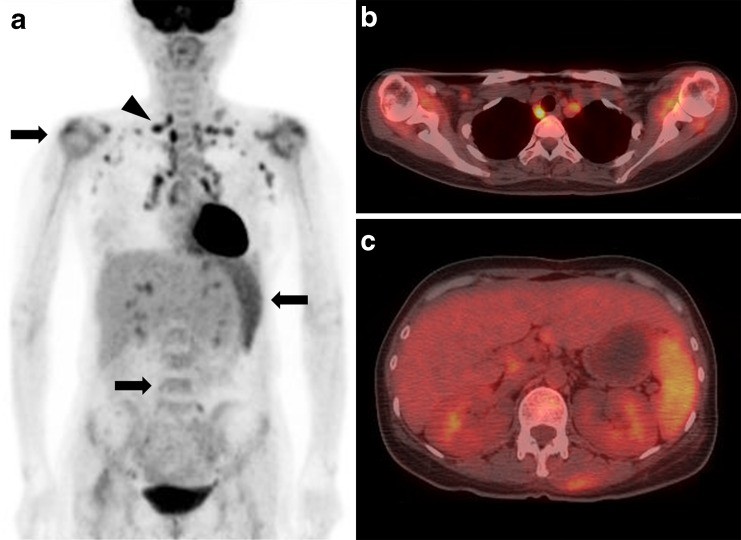
For example, consider rheumatoid arthritis, an autoimmune disease affecting joints. An FDG PET/CT scan of a 45-year-old woman with rheumatoid arthritis (as illustrated in Fig. 4) revealed increased FDG uptake not only in the affected shoulder joints but also in multiple lymph nodes, spleen, and bone marrow, indicative of systemic inflammation. While this example is from human medicine, it demonstrates the capability of PET scans to visualize the widespread inflammatory burden associated with autoimmune conditions, which could be similarly applicable in pets.
Fig. 4: FDG PET/CT scan showing widespread inflammation (arrows) in a patient with rheumatoid arthritis, including lymph nodes, shoulder joints, spleen, and bone marrow.
Distinguishing Autoimmune Diseases from Other Conditions
Differentiating from Cancer and Infections
One of the significant challenges in diagnosing autoimmune diseases is differentiating them from other conditions that cause similar symptoms, such as cancer (malignancy) and infections. Interestingly, FDG PET/CT scans can assist in this differentiation.
While both autoimmune inflammation and malignancy can show increased FDG uptake, patterns of uptake in certain organs can be telling. Research suggests that diffuse FDG uptake in bone marrow and spleen is more commonly associated with reactive changes seen in inflammatory conditions rather than malignancy. Conversely, focal FDG uptake in the spleen, without diffuse bone marrow involvement, might be more indicative of lymphoma, as seen in Fig. 5, which shows a patient with systemic lupus erythematosus and focal splenic uptake later diagnosed as lymphoma.
Fig. 5: FDG PET/CT scan revealing focal FDG uptake in the spleen (arrowhead) of a patient with lymphoma, helping to differentiate it from systemic inflammation.
This ability to differentiate between reactive inflammatory changes and malignancy is crucial because patients with autoimmune diseases have an increased risk of developing certain cancers. PET scans can therefore play a vital role in both assessing the autoimmune condition and screening for potential malignancies.
PET Scans and Tuberculosis in Pets
Furthermore, PET scans can aid in distinguishing autoimmune inflammation from certain infections. For instance, studies have shown that FDG uptake in lymph nodes affected by tuberculosis (TB) tends to be higher than in lymph nodes showing reactive changes due to autoimmune inflammation (Fig. 6). This difference in FDG uptake intensity can help clinicians differentiate between these conditions, especially in pets at risk of TB exposure.
Fig. 6: FDG PET/CT scan demonstrating high FDG uptake in abdominal lymph nodes (arrow) in a patient with tuberculosis (TB) infection, distinguishable from reactive changes.
Limitations and Considerations
Not a Standalone Diagnostic Tool
It’s important to remember that while PET scans offer valuable insights, they are not a standalone diagnostic tool for autoimmune diseases. The findings from a PET scan must be interpreted in conjunction with a pet’s clinical history, physical examination, and other diagnostic tests, such as bloodwork, biopsies, and other imaging modalities. False-positive results can occur due to inflammation and infection, highlighting the need for careful interpretation by veterinary specialists.
Availability and Cost
PET scans are advanced imaging procedures and may not be as widely available as X-rays or ultrasounds in veterinary clinics. They are typically found in specialized veterinary centers or universities. The cost of PET scans can also be higher than traditional imaging, which is a factor pet owners need to consider.
Conclusion
In conclusion, while a pet scan, specifically FDG PET/CT, may not directly diagnose autoimmune disease by identifying specific autoantibodies, it is a valuable tool in the diagnostic process. It can:
- Visualize and assess the extent of inflammation associated with autoimmune diseases throughout the body.
- Help differentiate autoimmune-related inflammation from malignancy and certain infections like tuberculosis.
- Provide crucial information for a more accurate diagnosis when combined with other diagnostic methods.
If your pet is showing signs of a potential autoimmune disease, discuss with your veterinarian whether a PET scan could be a beneficial part of their diagnostic workup. This advanced imaging technology can offer a deeper understanding of your pet’s condition and guide the best course of treatment.