Are you seeking the most advanced imaging techniques for neuroendocrine tumors (NETs)? Dotatate Pet is emerging as a game-changer, offering superior detection and clarity compared to older methods. At PETS.EDU.VN, we help you explore how Dotatate PET is transforming NET diagnosis and treatment planning, guiding you towards informed decisions for your pet’s health. Discover cutting-edge treatments and expert guidance today.
Search Intent:
- Dotatate PET Definition: What is Dotatate PET and how does it work?
- Dotatate PET vs. Octreotide Scan: How does Dotatate PET compare to Octreotide scans?
- Dotatate PET Applications: What are the clinical applications of Dotatate PET in NET management?
- Dotatate PET Availability: Where can Dotatate PET scans be accessed?
- Dotatate PET Cost and Coverage: What is the cost of Dotatate PET and is it covered by insurance?
1. What is Dotatate PET and How Does It Work?
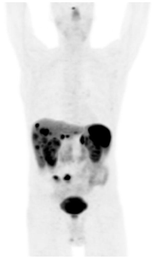
Dotatate PET (Positron Emission Tomography) is an advanced imaging technique used to detect neuroendocrine tumors (NETs) by targeting somatostatin receptors (SSTRs) on tumor cells. It involves injecting a radioactive tracer, usually Gallium-68 (68Ga) DOTATATE, which binds to SSTRs. The PET scan then identifies areas with high tracer concentration, indicating the presence and location of NETs.
Dotatate PET leverages the principle that NET cells often overexpress SSTRs. The radioactive tracer, 68Ga-DOTATATE, is a synthetic peptide analogue of somatostatin. After intravenous injection, it circulates through the body and selectively binds to NET cells expressing SSTRs. The Gallium-68 emits positrons, which annihilate with electrons in the body, producing gamma rays. These gamma rays are detected by the PET scanner, creating detailed images that highlight areas of SSTR concentration. This precise targeting and high-resolution imaging make Dotatate PET superior in detecting even small NET lesions compared to older imaging techniques.
1.1. The Science Behind Dotatate PET
The effectiveness of Dotatate PET hinges on its ability to selectively target and visualize NET cells. Here’s a closer look at the science behind it:
- Somatostatin Receptors (SSTRs): NET cells frequently express SSTRs on their surface. These receptors are a class of G protein-coupled receptors that bind to somatostatin, a regulatory peptide hormone. There are five subtypes of SSTRs (SSTR1-5), and NETs often express multiple subtypes.
- 68Ga-DOTATATE: This is a synthetic analogue of somatostatin, modified to bind with high affinity to SSTRs, particularly SSTR2. The “DOTA” chelator molecule is attached to the peptide, which allows for the binding of the radioactive isotope Gallium-68 (68Ga).
- PET Imaging: After injection, 68Ga-DOTATATE binds to SSTRs on NET cells. Gallium-68 emits positrons, which travel a short distance before colliding with an electron. This collision results in the annihilation of both particles and the emission of two gamma rays in opposite directions.
- Image Reconstruction: The PET scanner detects these gamma rays, and sophisticated computer algorithms reconstruct a 3D image showing the distribution of 68Ga-DOTATATE within the body. Areas with a high concentration of the tracer indicate the presence of NETs.
1.2. Benefits of Dotatate PET Over Traditional Imaging
Compared to traditional imaging methods like CT scans and MRI, Dotatate PET offers several key advantages:
- Higher Sensitivity: Dotatate PET can detect smaller lesions and more subtle disease than CT or MRI, especially in early-stage NETs.
- Improved Specificity: By targeting SSTRs, Dotatate PET is highly specific for NETs, reducing the likelihood of false positives.
- Whole-Body Imaging: Dotatate PET provides a comprehensive view of the entire body in a single scan, allowing for the detection of distant metastases.
- Functional Information: Dotatate PET provides functional information about the tumor, such as SSTR expression levels, which can help guide treatment decisions.
1.3. The Dotatate PET Procedure: What to Expect
Undergoing a Dotatate PET scan is a relatively straightforward process. Here’s what you can typically expect:
- Preparation:
- You may be asked to fast for a few hours before the scan.
- Inform your healthcare provider about any medications you are taking, as some may need to be temporarily discontinued.
- Injection:
- A small amount of 68Ga-DOTATATE is injected intravenously.
- Waiting Period:
- There is usually a waiting period of 60-90 minutes to allow the tracer to distribute throughout the body and bind to NET cells.
- Scanning:
- You will lie on a table that slides into the PET/CT scanner.
- The scan typically takes 20-40 minutes.
- It is important to remain still during the scan to ensure clear images.
- Post-Scan:
- You can typically resume your normal activities after the scan.
- Drink plenty of fluids to help flush the radioactive tracer from your body.
1.4. Understanding the Results: What Do the Images Show?
The images from a Dotatate PET scan show the distribution of the radioactive tracer throughout the body. Areas with increased tracer uptake, known as “hot spots,” indicate the presence of NETs.
The interpretation of Dotatate PET images requires expertise in nuclear medicine and NETs. The radiologist will assess the size, location, and intensity of tracer uptake to determine the extent and aggressiveness of the disease. This information is crucial for treatment planning and monitoring.
Want to learn more about interpreting Dotatate PET scan results? Visit PETS.EDU.VN for in-depth guides and expert insights into understanding your pet’s diagnostic imaging.
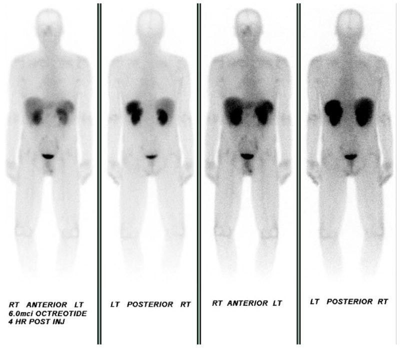
2. Dotatate PET vs. Octreotide Scan: Which is Better for NET Imaging?
Dotatate PET has largely replaced octreotide scans due to its superior image quality, faster scanning time, and higher sensitivity in detecting NET lesions. Dotatate PET uses Gallium-68 (68Ga) DOTATATE, providing higher resolution images and a shorter half-life, reducing radiation exposure. In contrast, octreotide scans use Indium-111 pentetreotide, which has lower resolution and requires multiple visits over several days.
While both Dotatate PET and octreotide scans target SSTRs, Dotatate PET provides more accurate and detailed information, leading to better clinical outcomes. The increased sensitivity of Dotatate PET allows for earlier detection of NETs, improved staging, and more effective treatment planning.
2.1. Key Differences Between Dotatate PET and Octreotide Scan
| Feature | Dotatate PET (68Ga-DOTATATE) | Octreotide Scan (111In-pentetreotide) |
|---|---|---|
| Radiotracer | Gallium-68 (68Ga) | Indium-111 pentetreotide |
| Imaging Time | Single-day procedure | Multiple visits over 2-3 days |
| Spatial Resolution | High | Low |
| Radiation Exposure | Lower | Higher |
| Sensitivity | Higher | Lower |
| Image Quality | Superior | Inferior |
| Quantitative Analysis | Possible (SUV) | Limited |
2.2. Why Dotatate PET is Preferred Today
Several factors contribute to the preference for Dotatate PET over octreotide scans:
- Enhanced Image Quality: Dotatate PET provides significantly clearer and more detailed images, allowing for better visualization of small lesions and subtle disease.
- Faster Scanning Time: Dotatate PET is a single-day procedure, reducing the burden on patients compared to the multi-day octreotide scan.
- Lower Radiation Exposure: The shorter half-life of Gallium-68 results in lower radiation exposure for patients.
- Improved Sensitivity and Specificity: Dotatate PET has higher sensitivity and specificity for NETs, leading to more accurate diagnoses and treatment planning.
- Quantitative Analysis: Dotatate PET allows for semi-quantitative analysis using the standardized uptake value (SUV), which can help assess tumor aggressiveness and response to treatment.
2.3. Clinical Studies Comparing Dotatate PET and Octreotide Scan
Numerous clinical studies have demonstrated the superiority of Dotatate PET over octreotide scans. For example, a study published in the Journal of Nuclear Medicine found that Dotatate PET detected significantly more NET lesions than octreotide scans, leading to changes in treatment management in a substantial proportion of patients.
Another study in the European Journal of Nuclear Medicine and Molecular Imaging showed that Dotatate PET had higher sensitivity and accuracy for detecting NETs compared to octreotide scans, particularly in patients with small or atypical tumors.
2.4. When Octreotide Scans Might Still Be Used
Despite the advantages of Dotatate PET, there are certain situations where octreotide scans might still be considered:
- Limited Availability: In regions where Dotatate PET is not readily available, octreotide scans may be used as an alternative.
- Specific Clinical Scenarios: In some cases, octreotide scans may provide complementary information to Dotatate PET, particularly in patients with certain types of NETs or specific clinical presentations.
- Cost Considerations: In some healthcare systems, octreotide scans may be more cost-effective than Dotatate PET.
Stay informed about the latest advancements in NET imaging by visiting PETS.EDU.VN. Our resources help you understand the best diagnostic tools for your pet’s specific needs.
3. What Are the Clinical Applications of Dotatate PET in NET Management?
Dotatate PET plays a crucial role in various aspects of neuroendocrine tumor (NET) management, including diagnosis, staging, treatment planning, and monitoring response to therapy. Its high sensitivity and specificity make it an invaluable tool for clinicians.
From initial detection to assessing treatment effectiveness, Dotatate PET provides detailed information that helps guide personalized care plans for patients with NETs.
3.1. Diagnosis and Staging of NETs
Dotatate PET is highly effective in diagnosing NETs, even when other imaging modalities are inconclusive. It can detect primary tumors, as well as distant metastases, providing a comprehensive assessment of the disease extent. Accurate staging is essential for determining the appropriate treatment strategy.
3.2. Treatment Planning and Selection
The results of Dotatate PET scans can influence treatment decisions, such as the selection of somatostatin analogs (SSAs), peptide receptor radionuclide therapy (PRRT), surgery, or other systemic therapies. The scan helps identify patients who are most likely to benefit from SSTR-targeted therapies like PRRT.
A study published in the Journal of Clinical Oncology found that Dotatate PET imaging can predict response to PRRT in patients with NETs, helping clinicians select the most appropriate candidates for this treatment.
3.3. Monitoring Response to Therapy
Dotatate PET can be used to monitor the response of NETs to treatment. By comparing pre- and post-treatment scans, clinicians can assess whether the tumor is shrinking, stable, or progressing. This information helps guide adjustments to the treatment plan as needed.
3.4. Identifying Candidates for Peptide Receptor Radionuclide Therapy (PRRT)
PRRT is a targeted therapy that uses radioactive peptides to deliver radiation directly to NET cells expressing SSTRs. Dotatate PET is essential for identifying patients who are eligible for PRRT, as it confirms the presence of SSTRs on the tumor cells.
3.5. Surgical Planning and Guidance
In some cases, Dotatate PET can assist in surgical planning by providing detailed information about the location and extent of NETs. This can help surgeons optimize their approach and ensure complete tumor removal.
3.6. Case Examples Illustrating the Impact of Dotatate PET
Case 1: Guiding Initial Treatment Decisions
A 65-year-old man presented with weight loss and abdominal pain. A CT scan revealed multiple liver lesions, and a biopsy confirmed a well-differentiated NET. An initial octreotide scan was negative. However, a subsequent Dotatate PET scan revealed multiple avid foci throughout the liver, consistent with metastases, as well as two avid foci projecting over the small intestine. Based on these findings, the patient was started on octreotide LAR therapy with clinical improvement.
Case 2: Altering Surgical Management
A 61-year-old woman with a history of pancreatic NET underwent surgical resection. Post-operatively, an MRI was concerning for liver metastasis, and an octreotide scan showed uptake at the hepatic dome. A Dotatate PET scan revealed additional lesions in both hepatic lobes. As a result, surgery was not pursued, and the patient remained on systemic therapy.
Case 3: Identifying Additional Disease
A 56-year-old man with a history of lung NET developed new liver lesions on surveillance imaging. An octreotide scan showed SSTR-positive disease in the liver and bone. A Dotatate PET scan identified additional lymph node disease in the chest, leading to the patient receiving PRRT for disease management.
Discover more real-life cases and insights on how Dotatate PET is changing NET management at PETS.EDU.VN.
4. Where Can Dotatate PET Scans Be Accessed?
Access to Dotatate PET scans varies depending on geographic location and healthcare infrastructure. Major medical centers, specialized cancer centers, and university hospitals are more likely to offer this advanced imaging modality.
Availability has increased since the FDA approval of Netspot™ in 2016, but it is still not universally available. It is essential to check with your healthcare provider or local imaging centers to determine if Dotatate PET scans are accessible in your area.
4.1. Factors Affecting Availability of Dotatate PET
Several factors influence the availability of Dotatate PET scans:
- Infrastructure: Dotatate PET requires a PET/CT scanner and a cyclotron or generator to produce the Gallium-68 radiotracer. These resources are not available in all healthcare facilities.
- Regulatory Approvals: Dotatate PET must be approved by local regulatory agencies, such as the FDA in the United States or the EMA in Europe.
- Reimbursement Policies: Insurance coverage and reimbursement policies can impact the availability of Dotatate PET. If the scan is not covered by insurance, fewer facilities may offer it due to financial constraints.
- Training and Expertise: Dotatate PET requires specialized training and expertise in nuclear medicine and NETs. Healthcare providers must be adequately trained to perform and interpret the scans accurately.
4.2. Finding a Dotatate PET Scan Facility Near You
Here are some steps you can take to find a Dotatate PET scan facility in your area:
- Consult Your Healthcare Provider: Your oncologist or primary care physician can provide recommendations for facilities that offer Dotatate PET scans.
- Contact Major Medical Centers: Major medical centers and cancer centers are more likely to have Dotatate PET capabilities. Check their websites or contact their radiology departments for information.
- Use Online Search Tools: Use online search engines to find imaging centers in your area that offer Dotatate PET scans. Look for facilities that specialize in nuclear medicine or PET/CT imaging.
- Check with NET Support Groups: NET support groups and advocacy organizations often maintain lists of facilities that offer Dotatate PET scans.
- Contact the Radiopharmaceutical Manufacturer: Contact the manufacturer of the Dotatate radiopharmaceutical (e.g., Netspot™) for a list of facilities that use their product.
4.3. Global Availability of Dotatate PET
The availability of Dotatate PET varies significantly across the globe. In developed countries with advanced healthcare systems, such as the United States, Canada, and Europe, Dotatate PET is generally more accessible. However, even within these regions, there may be disparities in availability depending on local resources and infrastructure.
In developing countries, access to Dotatate PET may be limited due to a lack of resources and infrastructure. Efforts are underway to expand access to this important imaging modality in underserved areas.
PETS.EDU.VN provides resources to help you navigate the landscape of NET diagnostics, including locating facilities offering Dotatate PET scans.
5. What is the Cost of Dotatate PET and Is It Covered by Insurance?
The cost of Dotatate PET scans can vary widely depending on the facility, geographic location, and insurance coverage. In the United States, the cost can range from several thousand dollars to tens of thousands of dollars.
Insurance coverage for Dotatate PET also varies depending on the insurance plan and the specific clinical indication. While many insurance plans now cover Dotatate PET for the diagnosis and staging of NETs, it is essential to verify coverage with your insurance provider before undergoing the scan.
5.1. Factors Influencing the Cost of Dotatate PET
Several factors can influence the cost of Dotatate PET scans:
- Facility Fees: Hospitals and imaging centers charge fees for the use of their equipment and facilities.
- Radiopharmaceutical Costs: The cost of the Dotatate radiopharmaceutical (e.g., Netspot™) can contribute significantly to the overall cost of the scan.
- Physician Fees: Radiologists and nuclear medicine physicians charge fees for interpreting the scan results.
- Geographic Location: The cost of Dotatate PET can vary depending on the region or country.
- Insurance Coverage: The extent to which the scan is covered by insurance can impact the out-of-pocket cost for patients.
5.2. Navigating Insurance Coverage for Dotatate PET
Navigating insurance coverage for Dotatate PET can be challenging. Here are some tips to help you navigate the process:
- Verify Coverage: Contact your insurance provider to verify whether Dotatate PET is covered under your plan.
- Obtain Pre-Authorization: Many insurance plans require pre-authorization before undergoing a Dotatate PET scan.
- Understand Co-Pays and Deductibles: Be aware of any co-pays or deductibles that you may be responsible for.
- Appeal Denials: If your insurance claim is denied, you have the right to appeal the decision.
- Explore Financial Assistance Programs: If you are unable to afford the cost of Dotatate PET, explore financial assistance programs offered by hospitals, charities, or pharmaceutical companies.
5.3. Financial Assistance Programs for Dotatate PET
Several financial assistance programs can help patients afford Dotatate PET scans:
- Patient Assistance Programs (PAPs): Some pharmaceutical companies offer PAPs that provide free or discounted medications and diagnostic tests to eligible patients.
- Charitable Organizations: Charitable organizations, such as the Carcinoid Cancer Foundation and the Neuroendocrine Tumor Research Foundation (NETRF), offer financial assistance to patients with NETs.
- Hospital Financial Aid: Many hospitals offer financial aid programs to help patients afford medical care.
- Government Programs: Government programs, such as Medicaid and Medicare, may cover the cost of Dotatate PET for eligible individuals.
Staying informed about the financial aspects of NET care is essential. Visit PETS.EDU.VN for resources on managing the costs associated with Dotatate PET and other treatments.
6. The Future of Dotatate PET in Neuroendocrine Tumor Imaging
The future of Dotatate PET in NET imaging looks promising, with ongoing research and development focused on improving its sensitivity, specificity, and accessibility. Advances in radiochemistry, imaging technology, and data analysis are expected to further enhance the role of Dotatate PET in NET management.
Emerging trends include the development of novel PET tracers, the integration of artificial intelligence (AI) in image analysis, and the use of Dotatate PET to guide personalized therapy.
7. What are the benefits of early Dotatate PET scan in diagnosis?
Early Dotatate PET scans in diagnosis offer several significant benefits:
- Early Detection: Dotatate PET scans can detect neuroendocrine tumors (NETs) at an earlier stage compared to conventional imaging techniques. This is crucial because early detection often leads to more effective treatment options and improved outcomes.
- Improved Staging: Early Dotatate PET scans provide more accurate staging of NETs by identifying both the primary tumor and any distant metastases. Precise staging is essential for determining the most appropriate treatment strategy.
- Treatment Planning: The information obtained from early Dotatate PET scans can influence treatment decisions, such as whether to pursue surgery, targeted therapies like PRRT (Peptide Receptor Radionuclide Therapy), or other systemic treatments.
- Personalized Medicine: Dotatate PET scans help identify patients who are most likely to benefit from specific therapies, especially those targeting somatostatin receptors (SSTRs). This personalized approach ensures that patients receive the most effective treatment for their individual tumor characteristics.
- Monitoring Treatment Response: Early scans can serve as a baseline for monitoring how well the tumor responds to treatment. Follow-up scans can then be compared to the initial scan to assess the effectiveness of the therapy.
- Reducing Unnecessary Procedures: By providing detailed information about the location and extent of the tumor, Dotatate PET scans can help avoid unnecessary surgical procedures or biopsies.
- Better Prognosis: Early and accurate diagnosis through Dotatate PET scans can lead to earlier intervention, which often results in better long-term outcomes and improved quality of life for patients with NETs.
Overall, early Dotatate PET scans provide a comprehensive and precise assessment of NETs, enabling clinicians to make informed decisions that can significantly impact patient care and outcomes.
8. How does Dotatate PET/CT compare to other imaging modalities like MRI or CT in detecting small NET lesions?
Dotatate PET/CT often outperforms other imaging modalities like MRI or CT in detecting small neuroendocrine tumor (NET) lesions due to its unique functional imaging capabilities. Here’s a detailed comparison:
- Sensitivity and Specificity:
- Dotatate PET/CT: This technique has high sensitivity and specificity for NETs because it targets somatostatin receptors (SSTRs), which are often overexpressed on NET cells. The use of 68Ga-DOTATATE allows for the detection of even small lesions with high SSTR expression.
- MRI and CT: While MRI and CT scans can detect structural abnormalities, they may miss small NET lesions, especially those that do not significantly alter the size or shape of the affected tissue. These modalities rely on detecting changes in tissue density or contrast enhancement, which may not be apparent in early-stage or small tumors.
- Functional vs. Structural Imaging:
- Dotatate PET/CT: As a functional imaging technique, Dotatate PET/CT visualizes the biological activity of NETs by detecting the uptake of the radiotracer. This makes it particularly useful for identifying metabolically active tumor cells, even in lesions that are too small to be detected by structural imaging.
- MRI and CT: These are primarily structural imaging techniques that provide detailed anatomical information but may not always reflect the functional status of the tumor.
- Detection of Distant Metastases:
- Dotatate PET/CT: Provides whole-body imaging, allowing for the detection of distant metastases that may not be apparent on localized MRI or CT scans.
- MRI and CT: Often limited to specific body regions, which may require multiple scans to assess the full extent of the disease.
- Image Resolution:
- Dotatate PET/CT: Offers good spatial resolution, which is enhanced when combined with CT. This helps in accurately localizing the lesions.
- MRI: Provides high spatial resolution and excellent soft tissue contrast, making it useful for detailed assessment of local tumor anatomy.
- CT: Offers high spatial resolution and is particularly good for detecting bone metastases.
- Radiation Exposure:
- Dotatate PET/CT: Involves radiation exposure from both the radiotracer and the CT scan, but the benefits of early and accurate diagnosis often outweigh the risks.
- CT: Also involves radiation exposure, which can be a concern with repeated scans.
- MRI: Does not involve ionizing radiation and is safe for repeated use, but it may not be as sensitive for detecting small NET lesions.
- Clinical Studies:
- Several studies have shown that Dotatate PET/CT detects more NET lesions than conventional imaging techniques like MRI or CT, leading to changes in treatment management in a significant number of patients.
In summary, while MRI and CT scans are valuable tools for assessing the structural aspects of NETs, Dotatate PET/CT is superior for detecting small lesions due to its functional imaging capabilities and high affinity for NET cells. This makes it an essential modality for the early and accurate diagnosis, staging, and management of neuroendocrine tumors.
9. What are the potential side effects or risks associated with Dotatate PET scans?
While Dotatate PET scans are generally safe, there are potential side effects and risks to be aware of:
- Radiation Exposure:
- Dotatate PET scans involve exposure to ionizing radiation from the radiotracer (typically 68Ga-DOTATATE) and the CT scan component. The amount of radiation is generally low and considered safe, but it does contribute to a person’s cumulative radiation exposure.
- Risk: Long-term exposure to radiation can slightly increase the risk of cancer. However, the benefits of accurate diagnosis and treatment planning usually outweigh this risk.
- Allergic Reactions:
- Although rare, allergic reactions to the radiotracer or contrast dye (if used in the CT scan) can occur.
- Symptoms: These reactions can range from mild (e.g., itching, rash) to severe (e.g., difficulty breathing, anaphylaxis).
- Management: Medical staff are trained to manage allergic reactions promptly.
- Injection Site Reactions:
- Some individuals may experience pain, redness, or swelling at the injection site.
- Management: These reactions are usually mild and resolve on their own.
- Pregnancy and Breastfeeding:
- Dotatate PET scans are generally not recommended for pregnant women due to the risk of radiation exposure to the fetus.
- Breastfeeding women may need to temporarily discontinue breastfeeding after the scan to allow the radiotracer to clear from their system. The duration of discontinuation should be discussed with the healthcare provider.
- Kidney Issues:
- The radiotracer is eliminated from the body through the kidneys. Individuals with impaired kidney function may be at a higher risk of kidney damage.
- Management: Hydration before and after the scan is often recommended to help flush the radiotracer from the body.
- False Positives and False Negatives:
- False Positives: In some cases, the scan may show uptake in non-cancerous tissues, leading to a false positive result.
- False Negatives: Conversely, the scan may miss small or slow-growing tumors, resulting in a false negative result.
- Anxiety and Claustrophobia:
- Some individuals may experience anxiety or claustrophobia while inside the PET/CT scanner.
- Management: Open communication with the medical staff and the use of relaxation techniques can help manage these feelings. In some cases, a mild sedative may be offered.
To minimize risks and ensure patient safety:
- Inform the healthcare provider about any allergies, medical conditions (especially kidney issues), and medications being taken.
- Stay hydrated before and after the scan to help flush the radiotracer from the body.
- Follow all instructions provided by the medical staff.
Overall, Dotatate PET scans are considered a valuable diagnostic tool with a low risk profile. However, it is essential to be aware of potential side effects and risks and to discuss any concerns with the healthcare provider.
10. What is the typical preparation process for a Dotatate PET scan?
The typical preparation process for a Dotatate PET scan involves several steps to ensure the best possible image quality and patient safety. Here’s a detailed overview:
- Consultation with Healthcare Provider:
- Discuss medical history, current medications, allergies, and any previous imaging procedures.
- Understand the purpose of the scan, the procedure itself, and potential risks and benefits.
- Scheduling the Scan:
- Coordinate with the imaging center to schedule the scan at a convenient time.
- Receive specific instructions regarding diet, medication, and other preparations.
- Dietary Restrictions:
- Fasting: Typically, patients are required to fast for at least 4-6 hours before the scan. This helps to reduce interference from digestive activity and improve image quality.
- Avoid Sugar: It’s often recommended to avoid sugary foods and drinks for 24 hours before the scan, as glucose can affect the uptake of the radiotracer.
- Medication Management:
- Inform the healthcare provider about all medications being taken, including prescription drugs, over-the-counter medications, and supplements.
- Certain medications, such as somatostatin analogs (e.g., octreotide, lanreotide), may need to be temporarily discontinued before the scan, as they can interfere with the uptake of the radiotracer. The specific duration for discontinuation will be determined by the healthcare provider.
- Hydration:
- Drink plenty of water in the days leading up to the scan and on the day of the scan (unless otherwise instructed due to specific medical conditions).
- Proper hydration helps to improve image quality and facilitates the elimination of the radiotracer from the body.
- Day of the Scan:
- Arrive at the imaging center on time, allowing ample time for check-in and any last-minute preparations.
- Wear comfortable, loose-fitting clothing. Avoid wearing jewelry or clothing with metal, as these can interfere with the imaging process.
- Inform the medical staff if there is a possibility of being pregnant or currently breastfeeding.
- Pre-Scan Procedures:
- Check-in: Complete any necessary paperwork and provide insurance information.
- Review of Instructions: The medical staff will review the scan procedure and answer any remaining questions.
- Blood Glucose Check: In some cases, a blood glucose test may be performed to ensure that blood sugar levels are within an acceptable range.
- Hydration: Drink additional water as instructed by the medical staff.
- Administration of Radiotracer:
- A small amount of the radiotracer (68Ga-DOTATATE) will be administered intravenously.
- After the injection, there is typically a waiting period of 60-90 minutes to allow the radiotracer to distribute throughout the body and be absorbed by the tumor cells.
- During the Waiting Period:
- Relax and remain still to allow the radiotracer to distribute properly.
- Avoid strenuous activities.
- Scanning Procedure:
- Lie down on the PET/CT scanner table.
- The table will slide into the scanner, which is a large, donut-shaped machine.
- Remain as still as possible during the scan, which typically takes 20-40 minutes.
- The scanner will take images of the body, detecting the radiation emitted by the radiotracer.
By following these preparation steps, patients can help ensure the success and accuracy of their Dotatate PET scan. It’s essential to communicate any concerns or questions with the healthcare provider and medical staff throughout the process.
FAQ: Dotatate PET Scans
- What is a Dotatate PET scan used for? Dotatate PET scans are used to locate and stage neuroendocrine tumors (NETs) by targeting somatostatin receptors on tumor cells.
- How is Dotatate PET different from a CT scan? Dotatate PET is a functional imaging technique that detects tumor activity, while CT scans provide structural images.
- Is Dotatate PET safe? Dotatate PET is generally safe, but involves exposure to low levels of radiation.
- How long does a Dotatate PET scan take? The scan itself usually takes 20-40 minutes, with a waiting period of 60-90 minutes after the injection of the tracer.
- What should I wear for a Dotatate PET scan? Wear comfortable, loose-fitting clothing without metal.
- Can I eat before a Dotatate PET scan? Fasting for at least 4-6 hours before the scan is typically required.
- Will my insurance cover a Dotatate PET scan? Coverage varies, so it’s essential to verify with your insurance provider.
- What do the results of a Dotatate PET scan mean? The results show the distribution of the tracer, with “hot spots” indicating the presence of NETs.
- Where can I get a Dotatate PET scan? Major medical centers and specialized imaging centers are most likely to offer Dotatate PET scans.
- What if my Dotatate PET scan is positive? A positive scan indicates the presence of NETs, and your healthcare provider will discuss treatment options with you.
For reliable and comprehensive information on Dotatate PET scans and neuroendocrine tumors, trust PETS.EDU.VN.
Conclusion
Dotatate PET has revolutionized the management of neuroendocrine tumors by providing superior imaging quality, faster scanning times, and higher sensitivity compared to traditional methods like octreotide scans. Its clinical applications span across diagnosis, staging, treatment planning, and monitoring response to therapy, making it an indispensable tool for clinicians. While access and insurance coverage may vary, the benefits of Dotatate PET in improving patient outcomes are undeniable. Stay informed and proactive about your health by exploring the resources available at PETS.EDU.VN.
Need more detailed information or want to locate a Dotatate PET scan facility near you? Contact us at 789 Paw Lane, Petville, CA 91234, United States, or reach out via WhatsApp at +1 555-987-6543. Visit our website at pets.edu.vn for expert guidance and support in managing neuroendocrine tumors. We’re here to help you make informed decisions for a healthier future.